 Movie
Movie Controller


+ Open data

- Basic information
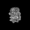
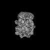
| Entry | Database: PDB / ID: 7y59 | |||||||||
|---|---|---|---|---|---|---|---|---|---|---|
| Title | The cryo-EM structure of human ERAD retro-translocation complex | |||||||||
 Components Components |
| |||||||||
 Keywords Keywords | MEMBRANE PROTEIN / ERAD | |||||||||
| Function / homology |  Function and homology information Function and homology informationDerlin-1-VIMP complex / signal recognition particle binding / endoplasmic reticulum quality control compartment / flavin adenine dinucleotide catabolic process / VCP-NSFL1C complex / endoplasmic reticulum stress-induced pre-emptive quality control / endosome to lysosome transport via multivesicular body sorting pathway / BAT3 complex binding / cytoplasmic ubiquitin ligase complex / cellular response to arsenite ion ...Derlin-1-VIMP complex / signal recognition particle binding / endoplasmic reticulum quality control compartment / flavin adenine dinucleotide catabolic process / VCP-NSFL1C complex / endoplasmic reticulum stress-induced pre-emptive quality control / endosome to lysosome transport via multivesicular body sorting pathway / BAT3 complex binding / cytoplasmic ubiquitin ligase complex / cellular response to arsenite ion / protein-DNA covalent cross-linking repair / Derlin-1 retrotranslocation complex / positive regulation of protein K63-linked deubiquitination / deubiquitinase activator activity / cytoplasm protein quality control / positive regulation of oxidative phosphorylation / aggresome assembly / ubiquitin-modified protein reader activity / regulation of protein localization to chromatin / mitotic spindle disassembly / VCP-NPL4-UFD1 AAA ATPase complex / cellular response to misfolded protein / positive regulation of mitochondrial membrane potential / vesicle-fusing ATPase / K48-linked polyubiquitin modification-dependent protein binding / NAD+ metabolic process / regulation of aerobic respiration / retrograde protein transport, ER to cytosol / stress granule disassembly / ATPase complex / ubiquitin-specific protease binding / regulation of synapse organization / ciliary transition zone / positive regulation of ATP biosynthetic process / intracellular membrane-bounded organelle / ubiquitin-like protein ligase binding / RHOH GTPase cycle / MHC class I protein binding / response to unfolded protein / autophagosome maturation / negative regulation of hippo signaling / HSF1 activation / endoplasmic reticulum to Golgi vesicle-mediated transport / polyubiquitin modification-dependent protein binding / interstrand cross-link repair / ATP metabolic process / translesion synthesis / Attachment and Entry / negative regulation of protein localization to chromatin / Protein methylation / endoplasmic reticulum unfolded protein response / ERAD pathway / proteasomal protein catabolic process / lipid droplet / ciliary tip / proteasome complex / viral genome replication / positive regulation of protein ubiquitination / Josephin domain DUBs / macroautophagy / negative regulation of smoothened signaling pathway / establishment of protein localization / N-glycan trimming in the ER and Calnexin/Calreticulin cycle / positive regulation of protein-containing complex assembly / Hh mutants are degraded by ERAD / ADP binding / Translesion Synthesis by POLH / Hedgehog ligand biogenesis / positive regulation of non-canonical NF-kappaB signal transduction / Defective CFTR causes cystic fibrosis / protein destabilization / autophagy / ABC-family protein mediated transport / cytoplasmic stress granule / Aggrephagy / positive regulation of protein catabolic process / azurophil granule lumen / Ovarian tumor domain proteases / late endosome / KEAP1-NFE2L2 pathway / positive regulation of canonical Wnt signaling pathway / double-strand break repair / positive regulation of proteasomal ubiquitin-dependent protein catabolic process / cellular response to heat / E3 ubiquitin ligases ubiquitinate target proteins / site of double-strand break / ATPase binding / signaling receptor activity / Neddylation / protease binding / secretory granule lumen / protein phosphatase binding / regulation of apoptotic process / ficolin-1-rich granule lumen / ubiquitin-dependent protein catabolic process / Attachment and Entry / proteasome-mediated ubiquitin-dependent protein catabolic process / early endosome / ciliary basal body / protein ubiquitination Similarity search - Function | |||||||||
| Biological species |  Homo sapiens (human) Homo sapiens (human) | |||||||||
| Method | ELECTRON MICROSCOPY / single particle reconstruction / cryo EM / Resolution: 4.51 Å | |||||||||
 Authors Authors | Cao, Y. / Rao, B. / Wang, Q. / Yao, D. / Xia, Y. / Li, W. / Li, S. / Shen, Y. | |||||||||
| Funding support |  China, 2items China, 2items
| |||||||||
 Citation Citation | Journal: Sci Adv / Year: 2023 Title: The cryo-EM structure of the human ERAD retrotranslocation complex. Authors: Bing Rao / Qian Wang / Deqiang Yao / Ying Xia / Wenguo Li / Yuming Xie / Shaobai Li / Mi Cao / Yafeng Shen / An Qin / Jie Zhao / Yu Cao / Abstract: Endoplasmic reticulum-associated degradation (ERAD) maintains protein homeostasis by retrieving misfolded proteins from the endoplasmic reticulum (ER) lumen into the cytosol for degradation. The ...Endoplasmic reticulum-associated degradation (ERAD) maintains protein homeostasis by retrieving misfolded proteins from the endoplasmic reticulum (ER) lumen into the cytosol for degradation. The retrotranslocation of misfolded proteins across the ER membrane is an energy-consuming process, with the detailed transportation mechanism still needing clarification. We determined the cryo-EM structures of the hetero-decameric complex formed by the Derlin-1 tetramer and the p97 hexamer. It showed an intriguing asymmetric complex and a putative coordinated squeezing movement in Derlin-1 and p97 parts. With the conformational changes of p97 induced by its ATP hydrolysis activities, the Derlin-1 channel could be torn into a "U" shape with a large opening to the lipidic environment, thereby forming an entry for the substrates in the ER membrane. The EM analysis showed that p97 formed a functional protein complex with Derlin-1, revealing the coupling mechanism between the ERAD retrotranslocation and the ATP hydrolysis activities. | |||||||||
| History |
|
- Structure visualization
| Structure viewer | Molecule: MolmilJmol/JSmol |
|---|
- Downloads & links
-Download
| PDBx/mmCIF format | 7y59.cif.gz | 904.3 KB | Display | PDBx/mmCIF format |
|---|---|---|---|---|
| PDB format | pdb7y59.ent.gz | 752 KB | Display | PDB format |
| PDBx/mmJSON format | 7y59.json.gz | Tree view | PDBx/mmJSON format | |
| Others |  Other downloads Other downloads |
-Validation report
| Arichive directory | https://data.pdbj.org/pub/pdb/validation_reports/y5/7y59ftp://data.pdbj.org/pub/pdb/validation_reports/y5/7y59 | HTTPS FTP |
|---|
-Related structure data
| Related structure data | 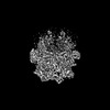 33613MC  7y4wC  7y53C M: map data used to model this data C: citing same article ( |
|---|---|
| Similar structure data |
-Links
 PDBj
PDBj


- Assembly
| Deposited unit | 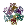
|
|---|---|
| 1 |
|
-Components
| #1: Protein | Mass: 30606.539 Da / Num. of mol.: 4 Source method: isolated from a genetically manipulated source Source: (gene. exp.) Homo sapiens (human) / Gene: DERL1, DER1, UNQ243/PRO276 / Cell line (production host): Sf9 / Production host:   Spodoptera frugiperda (fall armyworm) / References: UniProt: Q9BUN8 Spodoptera frugiperda (fall armyworm) / References: UniProt: Q9BUN8#2: Protein | Mass: 87546.711 Da / Num. of mol.: 6 Source method: isolated from a genetically manipulated source Source: (gene. exp.) Homo sapiens (human) / Gene: VCP / Cell line (production host): Sf9 / Production host: Spodoptera frugiperda (fall armyworm) / References: UniProt: P55072, vesicle-fusing ATPase#3: Chemical | ChemComp-ATP /   Mass: 507.181 Da / Num. of mol.: 6 / Source method: obtained synthetically / Formula: C10H16N5O13P3 / Feature type: SUBJECT OF INVESTIGATION / Comment: ATP, energy-carrying molecule*YM Mass: 507.181 Da / Num. of mol.: 6 / Source method: obtained synthetically / Formula: C10H16N5O13P3 / Feature type: SUBJECT OF INVESTIGATION / Comment: ATP, energy-carrying molecule*YM#4: Chemical | ChemComp-ADP /   Mass: 427.201 Da / Num. of mol.: 6 / Source method: obtained synthetically / Formula: C10H15N5O10P2 / Feature type: SUBJECT OF INVESTIGATION / Comment: ADP, energy-carrying molecule*YM Mass: 427.201 Da / Num. of mol.: 6 / Source method: obtained synthetically / Formula: C10H15N5O10P2 / Feature type: SUBJECT OF INVESTIGATION / Comment: ADP, energy-carrying molecule*YMHas ligand of interest | Y | |
|---|
-Experimental details
-Experiment
| Experiment | Method: ELECTRON MICROSCOPY |
|---|---|
| EM experiment | Aggregation state: PARTICLE / 3D reconstruction method: single particle reconstruction |
- Sample preparation
| Component | Name: The cryo-EM structure of human ERAD retro-translocation complex Type: COMPLEX / Entity ID: #1-#2 / Source: RECOMBINANT |
|---|---|
| Molecular weight | Value: 0.65 MDa / Experimental value: NO |
| Source (natural) | Organism: Homo sapiens (human) |
| Source (recombinant) | Organism: Spodoptera frugiperda (fall armyworm) |
| Buffer solution | pH: 7.5 |
| Specimen | Conc.: 15 mg/ml / Embedding applied: NO / Shadowing applied: NO / Staining applied: NO / Vitrification applied: YES |
| Specimen support | Grid material: GOLD / Grid mesh size: 300 divisions/in. / Grid type: Quantifoil R1.2/1.3 |
| Vitrification | Cryogen name: ETHANE / Humidity: 100 % / Chamber temperature: 281.15 K |
- Electron microscopy imaging
| Experimental equipment |  Model: Titan Krios / Image courtesy: FEI Company |
|---|---|
| Microscopy | Model: FEI TITAN KRIOS |
| Electron gun | Electron source:  FIELD EMISSION GUN / Accelerating voltage: 300 kV / Illumination mode: FLOOD BEAM FIELD EMISSION GUN / Accelerating voltage: 300 kV / Illumination mode: FLOOD BEAM |
| Electron lens | Mode: BRIGHT FIELD / Nominal magnification: 81000 X / Nominal defocus max: 2500 nm / Nominal defocus min: 1500 nm / Alignment procedure: COMA FREE |
| Specimen holder | Cryogen: NITROGEN / Specimen holder model: FEI TITAN KRIOS AUTOGRID HOLDER |
| Image recording | Average exposure time: 2.9 sec. / Electron dose: 50 e/Å2 / Film or detector model: GATAN K3 BIOQUANTUM (6k x 4k) |
| EM imaging optics | Energyfilter name: GIF Bioquantum / Energyfilter slit width: 15 eV |
- Processing
| EM software | Name: PHENIX / Version: 1.19.2_4158: / Category: model refinement | ||||||||||||||||||||||||
|---|---|---|---|---|---|---|---|---|---|---|---|---|---|---|---|---|---|---|---|---|---|---|---|---|---|
| CTF correction | Type: PHASE FLIPPING AND AMPLITUDE CORRECTION | ||||||||||||||||||||||||
| Symmetry | Point symmetry: C1 (asymmetric) | ||||||||||||||||||||||||
| 3D reconstruction | Resolution: 4.51 Å / Resolution method: FSC 0.143 CUT-OFF / Num. of particles: 62905 / Algorithm: FOURIER SPACE / Symmetry type: POINT | ||||||||||||||||||||||||
| Refine LS restraints |
|
