 Movie
Movie Controller


+ Open data

- Basic information
| Entry | Database: PDB / ID: 7y4r | ||||||
|---|---|---|---|---|---|---|---|
| Title | Structure of RclX | ||||||
 Components Components | CMD domain-containing protein | ||||||
 Keywords Keywords | UNKNOWN FUNCTION / peroxiredoxins / detoxification | ||||||
| Function / homology |  Function and homology information Function and homology information | ||||||
| Biological species |  Pseudomonas aeruginosa PAO1 (bacteria) Pseudomonas aeruginosa PAO1 (bacteria) | ||||||
| Method |  X-RAY DIFFRACTION / SYNCHROTRON / MOLECULAR REPLACEMENT / Resolution: 2.51 Å X-RAY DIFFRACTION / SYNCHROTRON / MOLECULAR REPLACEMENT / Resolution: 2.51 Å | ||||||
 Authors Authors | Ki, N. / Ha, N.C. | ||||||
| Funding support | 1items
| ||||||
 Citation Citation | Journal: To Be Published Title: Crystal structure of the putative HOCl and HOSCN-responsive peroxiredoxin RclX from Pseudomonas aeruginosa Authors: Ki, N. / Ha, N.C. | ||||||
| History |
|
- Structure visualization
| Structure viewer | Molecule: MolmilJmol/JSmol |
|---|
- Downloads & links
-Download
| PDBx/mmCIF format | 7y4r.cif.gz | 127.3 KB | Display | PDBx/mmCIF format |
|---|---|---|---|---|
| PDB format | pdb7y4r.ent.gz | 101.2 KB | Display | PDB format |
| PDBx/mmJSON format | 7y4r.json.gz | Tree view | PDBx/mmJSON format | |
| Others |  Other downloads Other downloads |
-Validation report
| Summary document | 7y4r_validation.pdf.gz | 488.2 KB | Display | wwPDB validaton report |
|---|---|---|---|---|
| Full document | 7y4r_full_validation.pdf.gz | 493.4 KB | Display | |
| Data in XML | 7y4r_validation.xml.gz | 22.9 KB | Display | |
| Data in CIF | 7y4r_validation.cif.gz | 32 KB | Display | |
| Arichive directory | https://data.pdbj.org/pub/pdb/validation_reports/y4/7y4rftp://data.pdbj.org/pub/pdb/validation_reports/y4/7y4r | HTTPS FTP |
-Related structure data
| Related structure data |  5dipS S: Starting model for refinement |
|---|---|
| Similar structure data |
-Links
 PDBj
PDBj- Assembly
| Deposited unit | 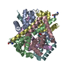
| ||||||||
|---|---|---|---|---|---|---|---|---|---|
| 1 |
| ||||||||
| Unit cell |
|
-Components
| #1: Protein | Mass: 11772.451 Da / Num. of mol.: 6 Source method: isolated from a genetically manipulated source Source: (gene. exp.) Pseudomonas aeruginosa PAO1 (bacteria)Strain: ATCC 15692 / DSM 22644 / CIP 104116 / JCM 14847 / LMG 12228 / 1C / PRS 101 / PAO1 Gene: PA0565 Production host: References: UniProt: Q9I5X1 #2: Chemical | ChemComp-PEO /   Mass: 34.015 Da / Num. of mol.: 5 / Source method: obtained synthetically / Formula: H2O2 Mass: 34.015 Da / Num. of mol.: 5 / Source method: obtained synthetically / Formula: H2O2#3: Chemical |   Type: L-peptide linking / Mass: 147.195 Da / Num. of mol.: 2 / Source method: isolated from a natural source / Formula: C6H15N2O2 Type: L-peptide linking / Mass: 147.195 Da / Num. of mol.: 2 / Source method: isolated from a natural source / Formula: C6H15N2O2#4: Water | ChemComp-HOH / |  Mass: 18.015 Da / Num. of mol.: 53 / Source method: isolated from a natural source / Formula: H2O Mass: 18.015 Da / Num. of mol.: 53 / Source method: isolated from a natural source / Formula: H2OHas ligand of interest | N | Has protein modification | Y | |
|---|
-Experimental details
-Experiment
| Experiment | Method: X-RAY DIFFRACTION / Number of used crystals: 1 |
|---|
- Sample preparation
| Crystal | Density Matthews: 2.48 Å3/Da / Density % sol: 50.43 % |
|---|---|
| Crystal grow | Temperature: 287.15 K / Method: vapor diffusion, hanging drop Details: 0.1 M sodium acetate trihydrate pH 4.6 15.5% (v/v) PEG 300 |
-Data collection
| Diffraction | Mean temperature: 100.15 K / Serial crystal experiment: N | |||||||||||||||||||||||||||||||||||||||||||||||||||||||||||||||||||||||||||||||||||||||||||||||||||||||||||||||||||||||||||||||||||||||||||||||||||||||||||||||||||||||||||||||||||||||||||||
|---|---|---|---|---|---|---|---|---|---|---|---|---|---|---|---|---|---|---|---|---|---|---|---|---|---|---|---|---|---|---|---|---|---|---|---|---|---|---|---|---|---|---|---|---|---|---|---|---|---|---|---|---|---|---|---|---|---|---|---|---|---|---|---|---|---|---|---|---|---|---|---|---|---|---|---|---|---|---|---|---|---|---|---|---|---|---|---|---|---|---|---|---|---|---|---|---|---|---|---|---|---|---|---|---|---|---|---|---|---|---|---|---|---|---|---|---|---|---|---|---|---|---|---|---|---|---|---|---|---|---|---|---|---|---|---|---|---|---|---|---|---|---|---|---|---|---|---|---|---|---|---|---|---|---|---|---|---|---|---|---|---|---|---|---|---|---|---|---|---|---|---|---|---|---|---|---|---|---|---|---|---|---|---|---|---|---|---|---|---|---|
| Diffraction source | Source: SYNCHROTRON / Site: PAL/PLS  / Beamline: 5C (4A) / Wavelength: 0.97942 Å / Beamline: 5C (4A) / Wavelength: 0.97942 Å | |||||||||||||||||||||||||||||||||||||||||||||||||||||||||||||||||||||||||||||||||||||||||||||||||||||||||||||||||||||||||||||||||||||||||||||||||||||||||||||||||||||||||||||||||||||||||||||
| Detector | Type: DECTRIS EIGER X 9M / Detector: PIXEL / Date: Nov 26, 2021 | |||||||||||||||||||||||||||||||||||||||||||||||||||||||||||||||||||||||||||||||||||||||||||||||||||||||||||||||||||||||||||||||||||||||||||||||||||||||||||||||||||||||||||||||||||||||||||||
| Radiation | Protocol: SINGLE WAVELENGTH / Monochromatic (M) / Laue (L): M / Scattering type: x-ray | |||||||||||||||||||||||||||||||||||||||||||||||||||||||||||||||||||||||||||||||||||||||||||||||||||||||||||||||||||||||||||||||||||||||||||||||||||||||||||||||||||||||||||||||||||||||||||||
| Radiation wavelength | Wavelength: 0.97942 Å / Relative weight: 1 | |||||||||||||||||||||||||||||||||||||||||||||||||||||||||||||||||||||||||||||||||||||||||||||||||||||||||||||||||||||||||||||||||||||||||||||||||||||||||||||||||||||||||||||||||||||||||||||
| Reflection | Resolution: 2.5→50 Å / Num. obs: 23999 / % possible obs: 96.7 % / Redundancy: 7.3 % / Biso Wilson estimate: 30.01 Å2 / Rmerge(I) obs: 0.143 / Rpim(I) all: 0.05 / Rrim(I) all: 0.152 / Χ2: 0.993 / Net I/σ(I): 6.5 / Num. measured all: 174870 | |||||||||||||||||||||||||||||||||||||||||||||||||||||||||||||||||||||||||||||||||||||||||||||||||||||||||||||||||||||||||||||||||||||||||||||||||||||||||||||||||||||||||||||||||||||||||||||
| Reflection shell | Diffraction-ID: 1
|
- Processing
| Software |
| |||||||||||||||||||||||||||||||||||||||||||||||||||||||||||||||
|---|---|---|---|---|---|---|---|---|---|---|---|---|---|---|---|---|---|---|---|---|---|---|---|---|---|---|---|---|---|---|---|---|---|---|---|---|---|---|---|---|---|---|---|---|---|---|---|---|---|---|---|---|---|---|---|---|---|---|---|---|---|---|---|---|
| Refinement | Method to determine structure: MOLECULAR REPLACEMENT Starting model: 5DIP Resolution: 2.51→37.61 Å / SU ML: 0.3 / Cross valid method: THROUGHOUT / σ(F): 2.23 / Phase error: 25.25 / Stereochemistry target values: ML
| |||||||||||||||||||||||||||||||||||||||||||||||||||||||||||||||
| Solvent computation | Shrinkage radii: 0.9 Å / VDW probe radii: 1.11 Å / Solvent model: FLAT BULK SOLVENT MODEL | |||||||||||||||||||||||||||||||||||||||||||||||||||||||||||||||
| Displacement parameters | Biso max: 95.09 Å2 / Biso mean: 31.8604 Å2 / Biso min: 5.89 Å2 | |||||||||||||||||||||||||||||||||||||||||||||||||||||||||||||||
| Refinement step | Cycle: final / Resolution: 2.51→37.61 Å
| |||||||||||||||||||||||||||||||||||||||||||||||||||||||||||||||
| LS refinement shell | Refine-ID: X-RAY DIFFRACTION / Rfactor Rfree error: 0 / Total num. of bins used: 8
|
