 Movie
Movie Controller


+ Open data

- Basic information
| Entry | Database: PDB / ID: 7y3x | |||||||||||||||
|---|---|---|---|---|---|---|---|---|---|---|---|---|---|---|---|---|
| Title | Crystal structure of BTG13 mutant (H58F) | |||||||||||||||
 Components Components | Questin oxidase | |||||||||||||||
 Keywords Keywords | OXIDOREDUCTASE / metalloenzyme / carboxylated-lysine | |||||||||||||||
| Function / homology | :  Function and homology information Function and homology information | |||||||||||||||
| Biological species |  Cercospora sojina (fungus) Cercospora sojina (fungus) | |||||||||||||||
| Method |  X-RAY DIFFRACTION / SYNCHROTRON / MOLECULAR REPLACEMENT / Resolution: 2.14 Å X-RAY DIFFRACTION / SYNCHROTRON / MOLECULAR REPLACEMENT / Resolution: 2.14 Å | |||||||||||||||
 Authors Authors | Hou, X.D. / Rao, Y.J. | |||||||||||||||
| Funding support |  China, 4items China, 4items
| |||||||||||||||
 Citation Citation | Journal: Angew.Chem.Int.Ed.Engl. / Year: 2022 Title: Discovery of the Biosynthetic Pathway of Beticolin 1 Reveals a Novel Non-Heme Iron-Dependent Oxygenase for Anthraquinone Ring Cleavage. Authors: Hou, X. / Xu, H. / Deng, Z. / Yan, Y. / Yuan, Z. / Liu, X. / Su, Z. / Yang, S. / Zhang, Y. / Rao, Y. | |||||||||||||||
| History |
|
- Structure visualization
| Structure viewer | Molecule: MolmilJmol/JSmol |
|---|
- Downloads & links
-Download
| PDBx/mmCIF format | 7y3x.cif.gz | 191.8 KB | Display | PDBx/mmCIF format |
|---|---|---|---|---|
| PDB format | pdb7y3x.ent.gz | 150 KB | Display | PDB format |
| PDBx/mmJSON format | 7y3x.json.gz | Tree view | PDBx/mmJSON format | |
| Others |  Other downloads Other downloads |
-Validation report
| Arichive directory | https://data.pdbj.org/pub/pdb/validation_reports/y3/7y3xftp://data.pdbj.org/pub/pdb/validation_reports/y3/7y3x | HTTPS FTP |
|---|
-Related structure data
| Related structure data |  7y3wSC 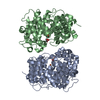 7y3yC S: Starting model for refinement C: citing same article ( |
|---|---|
| Similar structure data |
-Links
 PDBj
PDBj- Assembly
| Deposited unit | 
| ||||||||
|---|---|---|---|---|---|---|---|---|---|
| 1 | 
| ||||||||
| 2 | 
| ||||||||
| Unit cell |
|
-Components
| #1: Protein | Mass: 49453.094 Da / Num. of mol.: 2 / Mutation: H58F Source method: isolated from a genetically manipulated source Source: (gene. exp.) Cercospora sojina (fungus) / Production host:  #2: Chemical |   Mass: 55.845 Da / Num. of mol.: 2 / Source method: obtained synthetically / Formula: Fe / Feature type: SUBJECT OF INVESTIGATION Mass: 55.845 Da / Num. of mol.: 2 / Source method: obtained synthetically / Formula: Fe / Feature type: SUBJECT OF INVESTIGATION#3: Chemical |   Mass: 92.094 Da / Num. of mol.: 2 / Source method: obtained synthetically / Formula: C3H8O3 Mass: 92.094 Da / Num. of mol.: 2 / Source method: obtained synthetically / Formula: C3H8O3#4: Water | ChemComp-HOH / |  Mass: 18.015 Da / Num. of mol.: 419 / Source method: isolated from a natural source / Formula: H2O Mass: 18.015 Da / Num. of mol.: 419 / Source method: isolated from a natural source / Formula: H2OHas ligand of interest | Y | |
|---|
-Experimental details
-Experiment
| Experiment | Method: X-RAY DIFFRACTION / Number of used crystals: 1 |
|---|
- Sample preparation
| Crystal | Density Matthews: 2.59 Å3/Da / Density % sol: 52.46 % |
|---|---|
| Crystal grow | Temperature: 291 K / Method: vapor diffusion, sitting drop / Details: 0.1 M Bis-Tris pH 6.45, 16% PEG10000 |
-Data collection
| Diffraction | Mean temperature: 100 K / Serial crystal experiment: N | ||||||||||||||||||||||||||||||
|---|---|---|---|---|---|---|---|---|---|---|---|---|---|---|---|---|---|---|---|---|---|---|---|---|---|---|---|---|---|---|---|
| Diffraction source | Source: SYNCHROTRON / Site: NFPSS / Beamline: BL19U1 / Wavelength: 0.9785 Å | ||||||||||||||||||||||||||||||
| Detector | Type: DECTRIS PILATUS3 6M / Detector: PIXEL / Date: Nov 21, 2021 | ||||||||||||||||||||||||||||||
| Radiation | Protocol: SINGLE WAVELENGTH / Monochromatic (M) / Laue (L): M / Scattering type: x-ray | ||||||||||||||||||||||||||||||
| Radiation wavelength | Wavelength: 0.9785 Å / Relative weight: 1 | ||||||||||||||||||||||||||||||
| Reflection | Resolution: 2.14→79.01 Å / Num. obs: 57432 / % possible obs: 99.8 % / Redundancy: 13.2 % / CC1/2: 0.999 / Rmerge(I) obs: 0.14 / Rpim(I) all: 0.04 / Rrim(I) all: 0.146 / Net I/σ(I): 16.2 / Num. measured all: 757621 / Scaling rejects: 8 | ||||||||||||||||||||||||||||||
| Reflection shell | Diffraction-ID: 1
|
- Processing
| Software |
| ||||||||||||||||||||||||||||||||||||||||||||||||||||||||||||
|---|---|---|---|---|---|---|---|---|---|---|---|---|---|---|---|---|---|---|---|---|---|---|---|---|---|---|---|---|---|---|---|---|---|---|---|---|---|---|---|---|---|---|---|---|---|---|---|---|---|---|---|---|---|---|---|---|---|---|---|---|---|
| Refinement | Method to determine structure: MOLECULAR REPLACEMENT Starting model: 7Y3W Resolution: 2.14→79.01 Å / Cor.coef. Fo:Fc: 0.963 / Cor.coef. Fo:Fc free: 0.947 / SU B: 5.123 / SU ML: 0.126 / Cross valid method: THROUGHOUT / σ(F): 0 / ESU R: 0.204 / ESU R Free: 0.167 / Stereochemistry target values: MAXIMUM LIKELIHOOD Details: HYDROGENS HAVE BEEN ADDED IN THE RIDING POSITIONS U VALUES : REFINED INDIVIDUALLY
| ||||||||||||||||||||||||||||||||||||||||||||||||||||||||||||
| Solvent computation | Ion probe radii: 0.8 Å / Shrinkage radii: 0.8 Å / VDW probe radii: 1.2 Å / Solvent model: MASK | ||||||||||||||||||||||||||||||||||||||||||||||||||||||||||||
| Displacement parameters | Biso max: 105.71 Å2 / Biso mean: 34.43 Å2 / Biso min: 17.83 Å2
| ||||||||||||||||||||||||||||||||||||||||||||||||||||||||||||
| Refinement step | Cycle: final / Resolution: 2.14→79.01 Å
| ||||||||||||||||||||||||||||||||||||||||||||||||||||||||||||
| Refine LS restraints |
| ||||||||||||||||||||||||||||||||||||||||||||||||||||||||||||
| LS refinement shell | Resolution: 2.14→2.193 Å / Rfactor Rfree error: 0
|
