 Movie
Movie Controller

[English] 日本語
 Yorodumi
- PDB-7y1x: Crystal structure of prolyl oligopeptidase from Microbulbifer are... -
+ Open data

- Basic information
| Entry | Database: PDB / ID: 7y1x | ||||||
|---|---|---|---|---|---|---|---|
| Title | Crystal structure of prolyl oligopeptidase from Microbulbifer arenaceous complex with PEG400 and MES | ||||||
 Components Components | prolyl oligopeptidase | ||||||
 Keywords Keywords | HYDROLASE / S9A / prolyl endopeptidase / serine protease / mental disorder / amnesia | ||||||
| Function / homology | DI(HYDROXYETHYL)ETHER Function and homology information Function and homology information | ||||||
| Biological species |  Microbulbifer arenaceous (bacteria) Microbulbifer arenaceous (bacteria) | ||||||
| Method |  X-RAY DIFFRACTION / SYNCHROTRON / MOLECULAR REPLACEMENT / Resolution: 1.67 Å X-RAY DIFFRACTION / SYNCHROTRON / MOLECULAR REPLACEMENT / Resolution: 1.67 Å | ||||||
 Authors Authors | Huang, P. / Jiang, Z.Q. | ||||||
| Funding support |  China, 1items China, 1items
| ||||||
 Citation Citation | Journal: To Be Published Title: Crystal structure of prolyl oligopeptidase from Microbulbifer arenaceous complex with PEG400 and MES Authors: Huang, P. / Jiang, Z.Q. | ||||||
| History |
|
- Structure visualization
| Structure viewer | Molecule: MolmilJmol/JSmol |
|---|
- Downloads & links
-Download
| PDBx/mmCIF format | 7y1x.cif.gz | 167.1 KB | Display | PDBx/mmCIF format |
|---|---|---|---|---|
| PDB format | pdb7y1x.ent.gz | 124.3 KB | Display | PDB format |
| PDBx/mmJSON format | 7y1x.json.gz | Tree view | PDBx/mmJSON format | |
| Others |  Other downloads Other downloads |
-Validation report
| Summary document | 7y1x_validation.pdf.gz | 3 MB | Display | wwPDB validaton report |
|---|---|---|---|---|
| Full document | 7y1x_full_validation.pdf.gz | 3 MB | Display | |
| Data in XML | 7y1x_validation.xml.gz | 31.2 KB | Display | |
| Data in CIF | 7y1x_validation.cif.gz | 47.4 KB | Display | |
| Arichive directory | https://data.pdbj.org/pub/pdb/validation_reports/y1/7y1xftp://data.pdbj.org/pub/pdb/validation_reports/y1/7y1x | HTTPS FTP |
-Related structure data
| Related structure data | 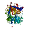 3munS S: Starting model for refinement |
|---|---|
| Similar structure data |
-Links
 PDBj
PDBj- Assembly
| Deposited unit | 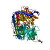
| ||||||||
|---|---|---|---|---|---|---|---|---|---|
| 1 |
| ||||||||
| Unit cell |
|
-Components
-Protein , 1 types, 1 molecules A
| #1: Protein | Mass: 79952.180 Da / Num. of mol.: 1 Source method: isolated from a genetically manipulated source Source: (gene. exp.) Microbulbifer arenaceous (bacteria) / Production host: |
|---|
-Non-polymers , 5 types, 531 molecules 


| #2: Chemical |  Mass: 238.278 Da / Num. of mol.: 2 / Source method: obtained synthetically / Formula: C10H22O6 / Feature type: SUBJECT OF INVESTIGATION / Comment: precipitant*YM Mass: 238.278 Da / Num. of mol.: 2 / Source method: obtained synthetically / Formula: C10H22O6 / Feature type: SUBJECT OF INVESTIGATION / Comment: precipitant*YM#3: Chemical | ChemComp-MES /  Mass: 195.237 Da / Num. of mol.: 7 / Source method: obtained synthetically / Formula: C6H13NO4S / Feature type: SUBJECT OF INVESTIGATION / Comment: pH buffer*YM Mass: 195.237 Da / Num. of mol.: 7 / Source method: obtained synthetically / Formula: C6H13NO4S / Feature type: SUBJECT OF INVESTIGATION / Comment: pH buffer*YM#4: Chemical | ChemComp-PEG / |  Mass: 106.120 Da / Num. of mol.: 1 / Source method: obtained synthetically / Formula: C4H10O3 / Feature type: SUBJECT OF INVESTIGATION Mass: 106.120 Da / Num. of mol.: 1 / Source method: obtained synthetically / Formula: C4H10O3 / Feature type: SUBJECT OF INVESTIGATION#5: Chemical | ChemComp-SO4 / |  Mass: 96.063 Da / Num. of mol.: 1 / Source method: obtained synthetically / Formula: SO4 / Feature type: SUBJECT OF INVESTIGATION Mass: 96.063 Da / Num. of mol.: 1 / Source method: obtained synthetically / Formula: SO4 / Feature type: SUBJECT OF INVESTIGATION#6: Water | ChemComp-HOH / | Mass: 18.015 Da / Num. of mol.: 520 / Source method: isolated from a natural source / Formula: H2O |
|---|
-Details
| Has ligand of interest | Y |
|---|---|
| Has protein modification | Y |
-Experimental details
-Experiment
| Experiment | Method: X-RAY DIFFRACTION / Number of used crystals: 1 |
|---|
- Sample preparation
| Crystal | Density Matthews: 2.19 Å3/Da / Density % sol: 43.88 % |
|---|---|
| Crystal grow | Temperature: 277 K / Method: vapor diffusion, hanging drop / pH: 6 Details: 0.04 M MES monohydrate pH 6.0, 8.8% (v/v) polyethylene glycol 400 |
-Data collection
| Diffraction | Mean temperature: 100 K / Serial crystal experiment: N |
|---|---|
| Diffraction source | Source: SYNCHROTRON / Site: SSRF / Beamline: BL17B1 / Wavelength: 0.97944 Å |
| Detector | Type: DECTRIS PILATUS3 S 2M / Detector: PIXEL / Date: Jun 27, 2021 |
| Radiation | Protocol: SINGLE WAVELENGTH / Monochromatic (M) / Laue (L): M / Scattering type: x-ray |
| Radiation wavelength | Wavelength: 0.97944 Å / Relative weight: 1 |
| Reflection | Resolution: 1.67→50 Å / Num. obs: 75931 / % possible obs: 94.38 % / Redundancy: 6.7 % / CC1/2: 0.998 / Net I/σ(I): 15.11 |
| Reflection shell | Resolution: 1.67→1.7 Å / Num. unique obs: 7149 / CC1/2: 0.899 |
- Processing
| Software |
| |||||||||||||||||||||||||||||||||||||||||||||||||||||||||||||||||||||||||||||||||||||||||||||||||||||||||||||||||||||||||||||||||||||||||||||||||||
|---|---|---|---|---|---|---|---|---|---|---|---|---|---|---|---|---|---|---|---|---|---|---|---|---|---|---|---|---|---|---|---|---|---|---|---|---|---|---|---|---|---|---|---|---|---|---|---|---|---|---|---|---|---|---|---|---|---|---|---|---|---|---|---|---|---|---|---|---|---|---|---|---|---|---|---|---|---|---|---|---|---|---|---|---|---|---|---|---|---|---|---|---|---|---|---|---|---|---|---|---|---|---|---|---|---|---|---|---|---|---|---|---|---|---|---|---|---|---|---|---|---|---|---|---|---|---|---|---|---|---|---|---|---|---|---|---|---|---|---|---|---|---|---|---|---|---|---|---|
| Refinement | Method to determine structure: MOLECULAR REPLACEMENT Starting model: 3MUN Resolution: 1.67→31.651 Å / Cor.coef. Fo:Fc: 0.96 / Cor.coef. Fo:Fc free: 0.951 / SU B: 1.705 / SU ML: 0.057 / Cross valid method: THROUGHOUT / ESU R: 0.099 / ESU R Free: 0.095 / Details: Hydrogens have not been used
| |||||||||||||||||||||||||||||||||||||||||||||||||||||||||||||||||||||||||||||||||||||||||||||||||||||||||||||||||||||||||||||||||||||||||||||||||||
| Solvent computation | Ion probe radii: 0.8 Å / Shrinkage radii: 0.8 Å / VDW probe radii: 1.2 Å / Solvent model: BABINET MODEL PLUS MASK | |||||||||||||||||||||||||||||||||||||||||||||||||||||||||||||||||||||||||||||||||||||||||||||||||||||||||||||||||||||||||||||||||||||||||||||||||||
| Displacement parameters | Biso mean: 16.037 Å2
| |||||||||||||||||||||||||||||||||||||||||||||||||||||||||||||||||||||||||||||||||||||||||||||||||||||||||||||||||||||||||||||||||||||||||||||||||||
| Refinement step | Cycle: LAST / Resolution: 1.67→31.651 Å
| |||||||||||||||||||||||||||||||||||||||||||||||||||||||||||||||||||||||||||||||||||||||||||||||||||||||||||||||||||||||||||||||||||||||||||||||||||
| Refine LS restraints |
| |||||||||||||||||||||||||||||||||||||||||||||||||||||||||||||||||||||||||||||||||||||||||||||||||||||||||||||||||||||||||||||||||||||||||||||||||||
| LS refinement shell |
|
