 Movie
Movie Controller

[English] 日本語
 Yorodumi
- PDB-7y17: Crystal structure of ribosomal ITS2 pre-rRNA processing complex f... -
+ Open data

- Basic information
| Entry | Database: PDB / ID: 7y17 | ||||||
|---|---|---|---|---|---|---|---|
| Title | Crystal structure of ribosomal ITS2 pre-rRNA processing complex from Cyberlindnera jadinii | ||||||
 Components Components |
| ||||||
 Keywords Keywords | RNA BINDING PROTEIN / RNA processing / Nuclease | ||||||
| Function / homology |  Function and homology information Function and homology informationpolynucleotide 5'-hydroxyl-kinase activity / Las1 complex / maturation of 5.8S rRNA / cleavage in ITS2 between 5.8S rRNA and LSU-rRNA of tricistronic rRNA transcript (SSU-rRNA, 5.8S rRNA, LSU-rRNA) / preribosome, large subunit precursor / maturation of LSU-rRNA / endonuclease activity / ATP binding / nucleus Similarity search - Function | ||||||
| Biological species |  Cyberlindnera jadinii (fungus) Cyberlindnera jadinii (fungus) | ||||||
| Method |  X-RAY DIFFRACTION / SYNCHROTRON / MOLECULAR REPLACEMENT / Resolution: 3.39 Å X-RAY DIFFRACTION / SYNCHROTRON / MOLECULAR REPLACEMENT / Resolution: 3.39 Å | ||||||
 Authors Authors | Chen, J. / Liu, L. | ||||||
| Funding support |  China, 1items China, 1items
| ||||||
 Citation Citation | Journal: Elife / Year: 2024 Title: Structural and mechanistic insights into ribosomal ITS2 RNA processing by nuclease-kinase machinery. Authors: Jiyun Chen / Hong Chen / Shanshan Li / Xiaofeng Lin / Rong Hu / Kaiming Zhang / Liang Liu / Abstract: Precursor ribosomal RNA (pre-rRNA) processing is a key step in ribosome biosynthesis and involves numerous RNases. A HEPN (higher eukaryote and prokaryote nucleotide binding) nuclease Las1 and a ...Precursor ribosomal RNA (pre-rRNA) processing is a key step in ribosome biosynthesis and involves numerous RNases. A HEPN (higher eukaryote and prokaryote nucleotide binding) nuclease Las1 and a polynucleotide kinase Grc3 assemble into a tetramerase responsible for rRNA maturation. Here, we report the structures of full-length and Las1-Grc3 complexes, and Las1. The Las1-Grc3 structures show that the central coiled-coil domain of Las1 facilitates pre-rRNA binding and cleavage, while the Grc3 C-terminal loop motif directly binds to the HEPN active center of Las1 and regulates pre-rRNA cleavage. Structural comparison between Las1 and Las1-Grc3 complex exhibits that Grc3 binding induces conformational rearrangements of catalytic residues associated with HEPN nuclease activation. Biochemical assays identify that Las1 processes pre-rRNA at the two specific sites (C2 and C2'), which greatly facilitates rRNA maturation. Our structures and specific pre-rRNA cleavage findings provide crucial insights into the mechanism and pathway of pre-rRNA processing in ribosome biosynthesis. | ||||||
| History |
|
- Structure visualization
| Structure viewer | Molecule: MolmilJmol/JSmol |
|---|
- Downloads & links
-Download
| PDBx/mmCIF format | 7y17.cif.gz | 514.5 KB | Display | PDBx/mmCIF format |
|---|---|---|---|---|
| PDB format | pdb7y17.ent.gz | 396.5 KB | Display | PDB format |
| PDBx/mmJSON format | 7y17.json.gz | Tree view | PDBx/mmJSON format | |
| Others |  Other downloads Other downloads |
-Validation report
| Arichive directory | https://data.pdbj.org/pub/pdb/validation_reports/y1/7y17ftp://data.pdbj.org/pub/pdb/validation_reports/y1/7y17 | HTTPS FTP |
|---|
-Related structure data
| Related structure data | 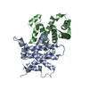 7y16C 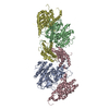 7y18C 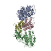 8j5yC 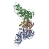 8j60C 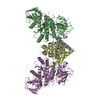 6of4S S: Starting model for refinement C: citing same article ( |
|---|---|
| Similar structure data |


-Links
 PDBj
PDBj- Assembly
| Deposited unit | 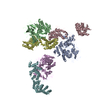
| ||||||||||||
|---|---|---|---|---|---|---|---|---|---|---|---|---|---|
| 1 | 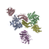
| ||||||||||||
| 2 | 
| ||||||||||||
| Unit cell |
| ||||||||||||
| Components on special symmetry positions |
|
-Components
| #1: Protein | Mass: 69612.867 Da / Num. of mol.: 3 Source method: isolated from a genetically manipulated source Source: (gene. exp.) Cyberlindnera jadinii (fungus)Strain: ATCC 18201 / CBS 1600 / BCRC 20928 / JCM 3617 / NBRC 0987 / NRRL Y-1542 Gene: GRC3, BN1211_2636, CYBJADRAFT_171070 / Production host:  #2: Protein | Mass: 49413.621 Da / Num. of mol.: 3 Source method: isolated from a genetically manipulated source Source: (gene. exp.) Cyberlindnera jadinii (fungus)Strain: ATCC 18201 / CBS 1600 / BCRC 20928 / JCM 3617 / NBRC 0987 / NRRL Y-1542 Gene: LAS1, BN1211_1791 / Production host: #3: Water | ChemComp-HOH / |  Mass: 18.015 Da / Num. of mol.: 386 / Source method: isolated from a natural source / Formula: H2O Mass: 18.015 Da / Num. of mol.: 386 / Source method: isolated from a natural source / Formula: H2OHas protein modification | Y | |
|---|
-Experimental details
-Experiment
| Experiment | Method: X-RAY DIFFRACTION / Number of used crystals: 1 |
|---|
- Sample preparation
| Crystal | Density Matthews: 3.04 Å3/Da / Density % sol: 59.53 % |
|---|---|
| Crystal grow | Temperature: 289 K / Method: vapor diffusion, hanging drop Details: 0.1 M sodium phosphate, pH 7.5, 0.05 M NaCl, and 9% PEG 4000 |
-Data collection
| Diffraction | Mean temperature: 100 K / Serial crystal experiment: N |
|---|---|
| Diffraction source | Source: SYNCHROTRON / Site: SSRF / Beamline: BL18U1 / Wavelength: 0.9793 Å |
| Detector | Type: DECTRIS PILATUS3 6M / Detector: PIXEL / Date: Jan 12, 2020 |
| Radiation | Protocol: SINGLE WAVELENGTH / Monochromatic (M) / Laue (L): M / Scattering type: x-ray |
| Radiation wavelength | Wavelength: 0.9793 Å / Relative weight: 1 |
| Reflection | Resolution: 3.23→50 Å / Num. obs: 41321 / % possible obs: 99.9 % / Redundancy: 7.5 % / Biso Wilson estimate: 71.33 Å2 / Rpim(I) all: 0.135 / Net I/σ(I): 4.3 |
| Reflection shell | Resolution: 3.23→3.29 Å / Num. unique obs: 3403 / Rpim(I) all: 0.412 |
- Processing
| Software |
| ||||||||||||||||||||||||||||||||||||||||||||||||||||||||||||||||||||||||||||||||||||||||||||||||||||||||||||||||
|---|---|---|---|---|---|---|---|---|---|---|---|---|---|---|---|---|---|---|---|---|---|---|---|---|---|---|---|---|---|---|---|---|---|---|---|---|---|---|---|---|---|---|---|---|---|---|---|---|---|---|---|---|---|---|---|---|---|---|---|---|---|---|---|---|---|---|---|---|---|---|---|---|---|---|---|---|---|---|---|---|---|---|---|---|---|---|---|---|---|---|---|---|---|---|---|---|---|---|---|---|---|---|---|---|---|---|---|---|---|---|---|---|---|
| Refinement | Method to determine structure: MOLECULAR REPLACEMENT Starting model: 6OF4 Resolution: 3.39→21.41 Å / SU ML: 0.6081 / Cross valid method: FREE R-VALUE / σ(F): 1.41 / Phase error: 31.8318 Stereochemistry target values: GeoStd + Monomer Library + CDL v1.2
| ||||||||||||||||||||||||||||||||||||||||||||||||||||||||||||||||||||||||||||||||||||||||||||||||||||||||||||||||
| Solvent computation | Shrinkage radii: 0.9 Å / VDW probe radii: 1.11 Å / Solvent model: FLAT BULK SOLVENT MODEL | ||||||||||||||||||||||||||||||||||||||||||||||||||||||||||||||||||||||||||||||||||||||||||||||||||||||||||||||||
| Displacement parameters | Biso mean: 124.13 Å2 | ||||||||||||||||||||||||||||||||||||||||||||||||||||||||||||||||||||||||||||||||||||||||||||||||||||||||||||||||
| Refinement step | Cycle: LAST / Resolution: 3.39→21.41 Å
| ||||||||||||||||||||||||||||||||||||||||||||||||||||||||||||||||||||||||||||||||||||||||||||||||||||||||||||||||
| Refine LS restraints |
| ||||||||||||||||||||||||||||||||||||||||||||||||||||||||||||||||||||||||||||||||||||||||||||||||||||||||||||||||
| LS refinement shell |
|
