 Movie
Movie Controller

[English] 日本語
 Yorodumi
- PDB-7y08: Crystal structure of Ricin A chain bound with (2-amino-4-oxo-3,4-... -
+ Open data

- Basic information
| Entry | Database: PDB / ID: 7y08 | ||||||
|---|---|---|---|---|---|---|---|
| Title | Crystal structure of Ricin A chain bound with (2-amino-4-oxo-3,4-dihydropteridine-7-carbonyl)glycyl-L-phenylalanine | ||||||
 Components Components | Ricin A chain | ||||||
 Keywords Keywords | HYDROLASE / HYDROLASE INHIBITOR | ||||||
| Function / homology |  Function and homology information Function and homology informationrRNA N-glycosylase / rRNA N-glycosylase activity / AMP binding / defense response / toxin activity / carbohydrate binding / killing of cells of another organism / negative regulation of translation Similarity search - Function | ||||||
| Biological species |  Ricinus communis (castor bean) Ricinus communis (castor bean) | ||||||
| Method |  X-RAY DIFFRACTION / SYNCHROTRON / MOLECULAR REPLACEMENT / molecular replacement / Resolution: 1.25 Å X-RAY DIFFRACTION / SYNCHROTRON / MOLECULAR REPLACEMENT / molecular replacement / Resolution: 1.25 Å | ||||||
 Authors Authors | Goto, M. / Higashi, S. / Ohba, T. / Kawata, R. / Nagatsu, K. / Suzuki, S. / Saito, R. | ||||||
| Funding support |  Japan, 1items Japan, 1items
| ||||||
 Citation Citation | Journal: Biochem.Biophys.Res.Commun. / Year: 2022 Title: Conformational change in ricin toxin A-Chain: A critical factor for inhibitor binding to the secondary pocket. Authors: Goto, M. / Higashi, S. / Ohba, T. / Kawata, R. / Nagatsu, K. / Suzuki, S. / Anslyn, E.V. / Saito, R. | ||||||
| History |
|
- Structure visualization
| Structure viewer | Molecule: MolmilJmol/JSmol |
|---|
- Downloads & links
-Download
| PDBx/mmCIF format | 7y08.cif.gz | 75.9 KB | Display | PDBx/mmCIF format |
|---|---|---|---|---|
| PDB format | pdb7y08.ent.gz | 52.6 KB | Display | PDB format |
| PDBx/mmJSON format | 7y08.json.gz | Tree view | PDBx/mmJSON format | |
| Others |  Other downloads Other downloads |
-Validation report
| Arichive directory | https://data.pdbj.org/pub/pdb/validation_reports/y0/7y08ftp://data.pdbj.org/pub/pdb/validation_reports/y0/7y08 | HTTPS FTP |
|---|
-Related structure data
| Related structure data |  7xzsC  7xztC 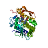 7xzuC 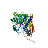 7xzwC 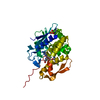 7y02C 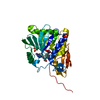 7y03C  7y05C  7y06C  7y07C  4huoS C: citing same article ( S: Starting model for refinement |
|---|---|
| Similar structure data |
-Links
 PDBj
PDBj

- Assembly
| Deposited unit | 
| ||||||||
|---|---|---|---|---|---|---|---|---|---|
| 1 |
| ||||||||
| Unit cell |
|
-Components
| #1: Protein | Mass: 30896.814 Da / Num. of mol.: 1 Source method: isolated from a genetically manipulated source Source: (gene. exp.) Ricinus communis (castor bean) / Plasmid: pET28a / Production host:  | ||||
|---|---|---|---|---|---|
| #2: Chemical | ChemComp-RS8 /   Type: peptide-like / Mass: 411.371 Da / Num. of mol.: 1 / Source method: obtained synthetically / Formula: C18H17N7O5 / Feature type: SUBJECT OF INVESTIGATION Type: peptide-like / Mass: 411.371 Da / Num. of mol.: 1 / Source method: obtained synthetically / Formula: C18H17N7O5 / Feature type: SUBJECT OF INVESTIGATION | ||||
| #3: Chemical |   Mass: 96.063 Da / Num. of mol.: 2 / Source method: obtained synthetically / Formula: SO4 Mass: 96.063 Da / Num. of mol.: 2 / Source method: obtained synthetically / Formula: SO4#4: Water | ChemComp-HOH / |  Mass: 18.015 Da / Num. of mol.: 303 / Source method: isolated from a natural source / Formula: H2O Mass: 18.015 Da / Num. of mol.: 303 / Source method: isolated from a natural source / Formula: H2OHas ligand of interest | Y | |
-Experimental details
-Experiment
| Experiment | Method: X-RAY DIFFRACTION / Number of used crystals: 1 |
|---|
- Sample preparation
| Crystal | Density Matthews: 2.58 Å3/Da / Density % sol: 52.38 % |
|---|---|
| Crystal grow | Temperature: 296 K / Method: vapor diffusion, hanging drop / pH: 5 / Details: Ammonium sulfate, Sodium malonate |
-Data collection
| Diffraction | Mean temperature: 95 K / Serial crystal experiment: N | |||||||||||||||||||||||||||||||||||||||||||||||||||||||||||||||||||||||||||||||||||||||||||||||||||||||||||||||||||||||||
|---|---|---|---|---|---|---|---|---|---|---|---|---|---|---|---|---|---|---|---|---|---|---|---|---|---|---|---|---|---|---|---|---|---|---|---|---|---|---|---|---|---|---|---|---|---|---|---|---|---|---|---|---|---|---|---|---|---|---|---|---|---|---|---|---|---|---|---|---|---|---|---|---|---|---|---|---|---|---|---|---|---|---|---|---|---|---|---|---|---|---|---|---|---|---|---|---|---|---|---|---|---|---|---|---|---|---|---|---|---|---|---|---|---|---|---|---|---|---|---|---|---|---|
| Diffraction source | Source: SYNCHROTRON / Site: Photon Factory / Beamline: AR-NW12A / Wavelength: 1 Å | |||||||||||||||||||||||||||||||||||||||||||||||||||||||||||||||||||||||||||||||||||||||||||||||||||||||||||||||||||||||||
| Detector | Type: ADSC QUANTUM 270 / Detector: CCD / Date: Nov 15, 2017 | |||||||||||||||||||||||||||||||||||||||||||||||||||||||||||||||||||||||||||||||||||||||||||||||||||||||||||||||||||||||||
| Radiation | Protocol: SINGLE WAVELENGTH / Monochromatic (M) / Laue (L): M / Scattering type: x-ray | |||||||||||||||||||||||||||||||||||||||||||||||||||||||||||||||||||||||||||||||||||||||||||||||||||||||||||||||||||||||||
| Radiation wavelength | Wavelength: 1 Å / Relative weight: 1 | |||||||||||||||||||||||||||||||||||||||||||||||||||||||||||||||||||||||||||||||||||||||||||||||||||||||||||||||||||||||||
| Reflection | Resolution: 1.25→60.729 Å / Num. all: 88699 / Num. obs: 88699 / % possible obs: 98.5 % / Redundancy: 13.4 % / Rpim(I) all: 0.015 / Rrim(I) all: 0.055 / Rsym value: 0.053 / Net I/av σ(I): 7.6 / Net I/σ(I): 29.2 / Num. measured all: 1188785 | |||||||||||||||||||||||||||||||||||||||||||||||||||||||||||||||||||||||||||||||||||||||||||||||||||||||||||||||||||||||||
| Reflection shell | Diffraction-ID: 1
|
-Phasing
| Phasing | Method: molecular replacement |
|---|
- Processing
| Software |
| |||||||||||||||||||||||||||||||||||||||||||||
|---|---|---|---|---|---|---|---|---|---|---|---|---|---|---|---|---|---|---|---|---|---|---|---|---|---|---|---|---|---|---|---|---|---|---|---|---|---|---|---|---|---|---|---|---|---|---|
| Refinement | Method to determine structure: MOLECULAR REPLACEMENT Starting model: 4HUO Resolution: 1.25→45.08 Å / Cor.coef. Fo:Fc: 0.938 / Cor.coef. Fo:Fc free: 0.926 / SU B: 0.753 / SU ML: 0.034 / SU R Cruickshank DPI: 0.0542 / Cross valid method: THROUGHOUT / σ(F): 0 / ESU R: 0.054 / ESU R Free: 0.055 / Stereochemistry target values: MAXIMUM LIKELIHOOD / Details: U VALUES : REFINED INDIVIDUALLY
| |||||||||||||||||||||||||||||||||||||||||||||
| Solvent computation | Ion probe radii: 0.8 Å / Shrinkage radii: 0.8 Å / VDW probe radii: 1.2 Å / Solvent model: MASK | |||||||||||||||||||||||||||||||||||||||||||||
| Displacement parameters | Biso max: 51.55 Å2 / Biso mean: 17.855 Å2 / Biso min: 9.09 Å2
| |||||||||||||||||||||||||||||||||||||||||||||
| Refinement step | Cycle: final / Resolution: 1.25→45.08 Å
| |||||||||||||||||||||||||||||||||||||||||||||
| Refine LS restraints |
| |||||||||||||||||||||||||||||||||||||||||||||
| LS refinement shell | Resolution: 1.25→1.282 Å / Rfactor Rfree error: 0 / Total num. of bins used: 20
|
