 Movie
Movie Controller


+ Open data

- Basic information
| Entry | Database: PDB / ID: 7xzo | ||||||
|---|---|---|---|---|---|---|---|
| Title | Formate-tetrahydrofolate ligase in complex with ATP | ||||||
 Components Components | Formate--tetrahydrofolate ligase | ||||||
 Keywords Keywords | LIGASE / Formate tetrahydrofolate ligase / ATP | ||||||
| Function / homology |  Function and homology information Function and homology informationformate-tetrahydrofolate ligase / formate-tetrahydrofolate ligase activity / tetrahydrofolate interconversion / ATP binding Similarity search - Function | ||||||
| Biological species |  Peptostreptococcus anaerobius (bacteria) Peptostreptococcus anaerobius (bacteria) | ||||||
| Method |  X-RAY DIFFRACTION / SYNCHROTRON / MOLECULAR REPLACEMENT / Resolution: 2.31 Å X-RAY DIFFRACTION / SYNCHROTRON / MOLECULAR REPLACEMENT / Resolution: 2.31 Å | ||||||
 Authors Authors | Fang, C.L. / Zhang, Y. | ||||||
| Funding support |  China, 1items China, 1items
| ||||||
 Citation Citation | Journal: Bmc Biol. / Year: 2023 Title: Identification of FtfL as a novel target of berberine in intestinal bacteria. Authors: Yan, J. / Fang, C. / Yang, G. / Li, J. / Liu, Y. / Zhang, L. / Yang, P. / Fang, J. / Gu, Y. / Zhang, Y. / Jiang, W. | ||||||
| History |
|
- Structure visualization
| Structure viewer | Molecule: MolmilJmol/JSmol |
|---|
- Downloads & links
-Download
| PDBx/mmCIF format | 7xzo.cif.gz | 421.3 KB | Display | PDBx/mmCIF format |
|---|---|---|---|---|
| PDB format | pdb7xzo.ent.gz | 343.1 KB | Display | PDB format |
| PDBx/mmJSON format | 7xzo.json.gz | Tree view | PDBx/mmJSON format | |
| Others |  Other downloads Other downloads |
-Validation report
| Arichive directory | https://data.pdbj.org/pub/pdb/validation_reports/xz/7xzoftp://data.pdbj.org/pub/pdb/validation_reports/xz/7xzo | HTTPS FTP |
|---|
-Related structure data
| Related structure data | 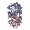 7xznC  7xzpC 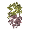 5a4jS S: Starting model for refinement C: citing same article ( |
|---|---|
| Similar structure data |
-Links
 PDBj
PDBj- Assembly
| Deposited unit | 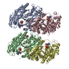
| ||||||||
|---|---|---|---|---|---|---|---|---|---|
| 1 | 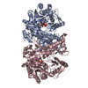
| ||||||||
| 2 | 
| ||||||||
| Unit cell |
|
-Components
| #1: Protein | Mass: 60583.137 Da / Num. of mol.: 4 Source method: isolated from a genetically manipulated source Source: (gene. exp.) Peptostreptococcus anaerobius (bacteria)Gene: fhs, NCTC11460_01517 / Production host: References: UniProt: A0A379CIH2, formate-tetrahydrofolate ligase #2: Chemical | ChemComp-K /   Mass: 39.098 Da / Num. of mol.: 4 / Source method: obtained synthetically / Formula: K Mass: 39.098 Da / Num. of mol.: 4 / Source method: obtained synthetically / Formula: K#3: Chemical | ChemComp-ATP /   Mass: 507.181 Da / Num. of mol.: 4 / Source method: obtained synthetically / Formula: C10H16N5O13P3 / Comment: ATP, energy-carrying molecule*YM Mass: 507.181 Da / Num. of mol.: 4 / Source method: obtained synthetically / Formula: C10H16N5O13P3 / Comment: ATP, energy-carrying molecule*YM#4: Chemical | ChemComp-BU3 / (   Mass: 90.121 Da / Num. of mol.: 5 / Source method: isolated from a natural source / Formula: C4H10O2 Mass: 90.121 Da / Num. of mol.: 5 / Source method: isolated from a natural source / Formula: C4H10O2#5: Water | ChemComp-HOH / |  Mass: 18.015 Da / Num. of mol.: 76 / Source method: isolated from a natural source / Formula: H2O Mass: 18.015 Da / Num. of mol.: 76 / Source method: isolated from a natural source / Formula: H2OHas ligand of interest | N | |
|---|
-Experimental details
-Experiment
| Experiment | Method: X-RAY DIFFRACTION / Number of used crystals: 1 |
|---|
- Sample preparation
| Crystal | Density Matthews: 2.72 Å3/Da / Density % sol: 54.76 % |
|---|---|
| Crystal grow | Temperature: 295.15 K / Method: vapor diffusion, hanging drop / Details: 20% w/v PEG 3350, 200mM potassium formate |
-Data collection
| Diffraction | Mean temperature: 100 K / Serial crystal experiment: N |
|---|---|
| Diffraction source | Source: SYNCHROTRON / Site: SSRF / Beamline: BL02U1 / Wavelength: 0.9791 Å |
| Detector | Type: DECTRIS EIGER2 S 9M / Detector: PIXEL / Date: Oct 10, 2021 |
| Radiation | Protocol: SINGLE WAVELENGTH / Monochromatic (M) / Laue (L): M / Scattering type: x-ray |
| Radiation wavelength | Wavelength: 0.9791 Å / Relative weight: 1 |
| Reflection | Resolution: 2.31→32.48 Å / Num. obs: 113515 / % possible obs: 99.9 % / Redundancy: 6.9 % / CC1/2: 0.995 / Net I/σ(I): 5.6 |
| Reflection shell | Resolution: 2.31→2.37 Å / Mean I/σ(I) obs: 1.2 / Num. unique obs: 8382 / CC1/2: 0.523 / % possible all: 99.9 |
- Processing
| Software |
| ||||||||||||||||||||||||||||||||||||||||||||||||||||||||||||||||||||||||||||||||||||||||||
|---|---|---|---|---|---|---|---|---|---|---|---|---|---|---|---|---|---|---|---|---|---|---|---|---|---|---|---|---|---|---|---|---|---|---|---|---|---|---|---|---|---|---|---|---|---|---|---|---|---|---|---|---|---|---|---|---|---|---|---|---|---|---|---|---|---|---|---|---|---|---|---|---|---|---|---|---|---|---|---|---|---|---|---|---|---|---|---|---|---|---|---|
| Refinement | Method to determine structure: MOLECULAR REPLACEMENT Starting model: 5a4j Resolution: 2.31→30.029 Å / SU ML: 0.35 / Cross valid method: THROUGHOUT / σ(F): 1.4 / Phase error: 34.9 / Stereochemistry target values: ML
| ||||||||||||||||||||||||||||||||||||||||||||||||||||||||||||||||||||||||||||||||||||||||||
| Solvent computation | Shrinkage radii: 0.9 Å / VDW probe radii: 1.11 Å / Solvent model: FLAT BULK SOLVENT MODEL | ||||||||||||||||||||||||||||||||||||||||||||||||||||||||||||||||||||||||||||||||||||||||||
| Displacement parameters | Biso max: 104.84 Å2 / Biso mean: 42.4203 Å2 / Biso min: 13.57 Å2 | ||||||||||||||||||||||||||||||||||||||||||||||||||||||||||||||||||||||||||||||||||||||||||
| Refinement step | Cycle: final / Resolution: 2.31→30.029 Å
| ||||||||||||||||||||||||||||||||||||||||||||||||||||||||||||||||||||||||||||||||||||||||||
| LS refinement shell | Refine-ID: X-RAY DIFFRACTION / Rfactor Rfree error: 0
|
