 Movie
Movie Controller


+ Open data

- Basic information
| Entry | Database: PDB / ID: 7xwi | ||||||
|---|---|---|---|---|---|---|---|
| Title | structure of patulin-detoxifying enzyme with NADPH | ||||||
 Components Components | Short-chain dehydrogenase/reductase | ||||||
 Keywords Keywords | OXIDOREDUCTASE / dehydrogenases/reductase | ||||||
| Function / homology | : / alcohol dehydrogenase / oxidoreductase activity, acting on the CH-OH group of donors, NAD or NADP as acceptor / Enoyl-(Acyl carrier protein) reductase / Short-chain dehydrogenase/reductase SDR / NAD(P)-binding domain superfamily / nucleotide binding / Chem-NDP / Short-chain dehydrogenase/reductase Function and homology information Function and homology information | ||||||
| Biological species |  Meyerozyma guilliermondii (fungus) Meyerozyma guilliermondii (fungus) | ||||||
| Method |  X-RAY DIFFRACTION / MOLECULAR REPLACEMENT / Resolution: 2.22 Å X-RAY DIFFRACTION / MOLECULAR REPLACEMENT / Resolution: 2.22 Å | ||||||
 Authors Authors | Dai, L. / Li, H. / Hu, Y. / Guo, R.T. / Chen, C.C. | ||||||
| Funding support | 1items
| ||||||
 Citation Citation | Journal: Int.J.Biol.Macromol. / Year: 2022 Title: Structure-based rational design of a short-chain dehydrogenase/reductase for improving activity toward mycotoxin patulin. Authors: Dai, L. / Li, H. / Huang, J.W. / Hu, Y. / He, M. / Yang, Y. / Min, J. / Guo, R.T. / Chen, C.C. | ||||||
| History |
|
- Structure visualization
| Structure viewer | Molecule: MolmilJmol/JSmol |
|---|
- Downloads & links
-Download
| PDBx/mmCIF format | 7xwi.cif.gz | 199.5 KB | Display | PDBx/mmCIF format |
|---|---|---|---|---|
| PDB format | pdb7xwi.ent.gz | 158.2 KB | Display | PDB format |
| PDBx/mmJSON format | 7xwi.json.gz | Tree view | PDBx/mmJSON format | |
| Others |  Other downloads Other downloads |
-Validation report
| Summary document | 7xwi_validation.pdf.gz | 1.6 MB | Display | wwPDB validaton report |
|---|---|---|---|---|
| Full document | 7xwi_full_validation.pdf.gz | 1.6 MB | Display | |
| Data in XML | 7xwi_validation.xml.gz | 42 KB | Display | |
| Data in CIF | 7xwi_validation.cif.gz | 59 KB | Display | |
| Arichive directory | https://data.pdbj.org/pub/pdb/validation_reports/xw/7xwiftp://data.pdbj.org/pub/pdb/validation_reports/xw/7xwi | HTTPS FTP |
-Related structure data
| Related structure data |  7xwhSC  7xwjC 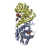 7xwkC 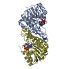 7xwlC 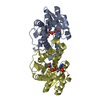 7xwmC 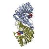 7xwnC S: Starting model for refinement C: citing same article ( |
|---|---|
| Similar structure data |
-Links
 PDBj
PDBj


- Assembly
| Deposited unit | 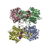
| ||||||||
|---|---|---|---|---|---|---|---|---|---|
| 1 | 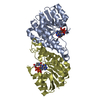
| ||||||||
| 2 | 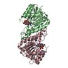
| ||||||||
| Unit cell |
|
-Components
| #1: Protein | Mass: 28070.781 Da / Num. of mol.: 4 Source method: isolated from a genetically manipulated source Source: (gene. exp.) Meyerozyma guilliermondii (fungus) / Gene: SDR / Production host:  #2: Chemical | ChemComp-NDP /   Mass: 745.421 Da / Num. of mol.: 4 / Source method: obtained synthetically / Formula: C21H30N7O17P3 / Feature type: SUBJECT OF INVESTIGATION Mass: 745.421 Da / Num. of mol.: 4 / Source method: obtained synthetically / Formula: C21H30N7O17P3 / Feature type: SUBJECT OF INVESTIGATION#3: Water | ChemComp-HOH / |  Mass: 18.015 Da / Num. of mol.: 595 / Source method: isolated from a natural source / Formula: H2O Mass: 18.015 Da / Num. of mol.: 595 / Source method: isolated from a natural source / Formula: H2OHas ligand of interest | Y | |
|---|
-Experimental details
-Experiment
| Experiment | Method: X-RAY DIFFRACTION / Number of used crystals: 1 |
|---|
- Sample preparation
| Crystal | Density Matthews: 2.08 Å3/Da / Density % sol: 40.93 % |
|---|---|
| Crystal grow | Temperature: 298 K / Method: vapor diffusion, sitting drop / Details: PEG Smear Broad, 0.1 M MES pH 6.5 |
-Data collection
| Diffraction | Mean temperature: 100 K / Serial crystal experiment: N |
|---|---|
| Diffraction source | Source: LIQUID ANODE / Type: BRUKER METALJET / Wavelength: 1.34138 Å |
| Detector | Type: BRUKER PHOTON 100 / Detector: CMOS / Date: Jul 9, 2021 |
| Radiation | Protocol: SINGLE WAVELENGTH / Monochromatic (M) / Laue (L): M / Scattering type: x-ray |
| Radiation wavelength | Wavelength: 1.34138 Å / Relative weight: 1 |
| Reflection | Resolution: 2.22→34.96 Å / Num. obs: 45943 / % possible obs: 99.9 % / Redundancy: 5.98 % / Rmerge(I) obs: 0.1528 / Net I/σ(I): 6.9 |
| Reflection shell | Resolution: 2.22→2.25 Å / Rmerge(I) obs: 0.484 / Num. unique obs: 1834 |
- Processing
| Software |
| ||||||||||||||||||||||||||||||||||||||||||||||||||||||||||||
|---|---|---|---|---|---|---|---|---|---|---|---|---|---|---|---|---|---|---|---|---|---|---|---|---|---|---|---|---|---|---|---|---|---|---|---|---|---|---|---|---|---|---|---|---|---|---|---|---|---|---|---|---|---|---|---|---|---|---|---|---|---|
| Refinement | Method to determine structure: MOLECULAR REPLACEMENT Starting model: 7XWH Resolution: 2.22→34.96 Å / Cor.coef. Fo:Fc: 0.941 / Cor.coef. Fo:Fc free: 0.883 / SU B: 8.742 / SU ML: 0.215 / SU R Cruickshank DPI: 0.3476 / Cross valid method: THROUGHOUT / σ(F): 0 / ESU R: 0.348 / ESU R Free: 0.263 / Stereochemistry target values: MAXIMUM LIKELIHOOD Details: HYDROGENS HAVE BEEN ADDED IN THE RIDING POSITIONS U VALUES : REFINED INDIVIDUALLY
| ||||||||||||||||||||||||||||||||||||||||||||||||||||||||||||
| Solvent computation | Ion probe radii: 0.8 Å / Shrinkage radii: 0.8 Å / VDW probe radii: 1.2 Å / Solvent model: MASK | ||||||||||||||||||||||||||||||||||||||||||||||||||||||||||||
| Displacement parameters | Biso max: 122.73 Å2 / Biso mean: 26.838 Å2 / Biso min: 9.2 Å2
| ||||||||||||||||||||||||||||||||||||||||||||||||||||||||||||
| Refinement step | Cycle: final / Resolution: 2.22→34.96 Å
| ||||||||||||||||||||||||||||||||||||||||||||||||||||||||||||
| Refine LS restraints |
| ||||||||||||||||||||||||||||||||||||||||||||||||||||||||||||
| LS refinement shell | Resolution: 2.22→2.278 Å / Rfactor Rfree error: 0 / Total num. of bins used: 20
|
