| Entry | Database: PDB / ID: 7xu8
|
|---|
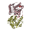
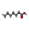
| Title | Structure of the complex of camel peptidoglycan recognition protein-short (PGRP-S) with heptanoic acid at 2.15 A resolution. |
|---|
 Components Components | Peptidoglycan recognition protein 1 |
|---|
 Keywords Keywords | IMMUNE SYSTEM / CPGRP-S / innate immune system |
|---|
| Function / homology |  Function and homology information Function and homology information
peptidoglycan immune receptor activity / N-acetylmuramoyl-L-alanine amidase activity / peptidoglycan binding / negative regulation of cytokine production / detection of bacterium / peptidoglycan catabolic process / defense response to Gram-positive bacterium / innate immune response / extracellular region / zinc ion bindingSimilarity search - Function Peptidoglycan recognition protein, PGRP-S / Peptidoglycan recognition protein family domain, metazoa/bacteria / Peptidoglycan recognition protein / Animal peptidoglycan recognition proteins homologous to Bacteriophage T3 lysozyme. / N-acetylmuramoyl-L-alanine amidase / Ami_2 / N-acetylmuramoyl-L-alanine amidase domain / N-acetylmuramoyl-L-alanine amidase/PGRP domain superfamilySimilarity search - Domain/homology |
|---|
| Biological species |   Camelus dromedarius (Arabian camel) Camelus dromedarius (Arabian camel) |
|---|
| Method |  X-RAY DIFFRACTION / SYNCHROTRON / MOLECULAR REPLACEMENT / Resolution: 2.15 Å X-RAY DIFFRACTION / SYNCHROTRON / MOLECULAR REPLACEMENT / Resolution: 2.15 Å |
|---|
 Authors Authors | Maurya, A. / Ahmad, N. / Viswanathan, V. / Singh, P.K. / Yamini, S. / Sharma, P. / Sinha, M. / Bhushan, A. / Kaur, P. / Sharma, S. / Singh, T.P. |
|---|
| Funding support |  India, 1items India, 1items | Organization | Grant number | Country |
|---|
| Not funded | | India |
|
|---|
 Citation Citation | Journal: Int J Biochem Mol Biol / Year: 2022
Title: Ligand recognition by peptidoglycan recognition protein-S (PGRP-S): structure of the complex of camel PGRP-S with heptanoic acid at 2.15 angstrom resolution.
Authors: Maurya, A. / Ahmad, N. / Singh, P.K. / Viswanathan, V. / Kaur, P. / Sharma, P. / Sharma, S. / Singh, T.P. |
|---|
| History | | Deposition | May 18, 2022 | Deposition site: PDBJ / Processing site: PDBJ |
|---|
| Supersession | Jun 15, 2022 | ID: 3UML |
|---|
| Revision 1.0 | Jun 15, 2022 | Provider: repository / Type: Initial release |
|---|
| Revision 1.1 | Oct 12, 2022 | Group: Database references / Derived calculations / Category: atom_type / citation / citation_author
Item: _atom_type.pdbx_N_electrons / _atom_type.pdbx_scat_Z ..._atom_type.pdbx_N_electrons / _atom_type.pdbx_scat_Z / _citation.country / _citation.journal_abbrev / _citation.journal_id_CSD / _citation.journal_id_ISSN / _citation.journal_volume / _citation.page_first / _citation.page_last / _citation.pdbx_database_id_PubMed / _citation.title / _citation.year |
|---|
| Revision 1.2 | Nov 29, 2023 | Group: Data collection / Refinement description
Category: chem_comp_atom / chem_comp_bond ...chem_comp_atom / chem_comp_bond / pdbx_initial_refinement_model / struct_ncs_dom_lim
Item: _struct_ncs_dom_lim.beg_auth_comp_id / _struct_ncs_dom_lim.beg_label_asym_id ..._struct_ncs_dom_lim.beg_auth_comp_id / _struct_ncs_dom_lim.beg_label_asym_id / _struct_ncs_dom_lim.beg_label_comp_id / _struct_ncs_dom_lim.beg_label_seq_id / _struct_ncs_dom_lim.end_auth_comp_id / _struct_ncs_dom_lim.end_label_asym_id / _struct_ncs_dom_lim.end_label_comp_id / _struct_ncs_dom_lim.end_label_seq_id |
|---|
| Revision 1.3 | Oct 16, 2024 | Group: Structure summary / Category: pdbx_entry_details / pdbx_modification_feature / Item: _pdbx_entry_details.has_protein_modification |
|---|
|
|---|
 Movie
Movie Controller

 Yorodumi
 Open data
 Basic information
 Structure visualization
 Downloads & links
 Other downloads
 PDBj
PDBj


 Assembly
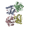


 Mass: 62.068 Da / Num. of mol.: 4 / Source method: obtained synthetically / Formula: C2H6O2 / Feature type: SUBJECT OF INVESTIGATION
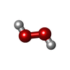
 Mass: 34.015 Da / Num. of mol.: 9 / Source method: obtained synthetically / Formula: H2O2 / Feature type: SUBJECT OF INVESTIGATION
 Mass: 60.009 Da / Num. of mol.: 1 / Source method: obtained synthetically / Formula: CO3 / Feature type: SUBJECT OF INVESTIGATION
 Mass: 22.990 Da / Num. of mol.: 1 / Source method: obtained synthetically / Formula: Na / Feature type: SUBJECT OF INVESTIGATION
 Mass: 130.185 Da / Num. of mol.: 1 / Source method: obtained synthetically / Formula: C7H14O2 / Feature type: SUBJECT OF INVESTIGATION
 Mass: 118.174 Da / Num. of mol.: 1 / Source method: obtained synthetically / Formula: C6H14O2 / Feature type: SUBJECT OF INVESTIGATION / Comment: precipitant*YM
 Sample preparation
 / Beamline: BM14 / Wavelength: 0.97 Å
 Processing