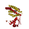
 Movie
Movie Controller


+ Open data

- Basic information
| Entry | Database: PDB / ID: 7xp0 | ||||||
|---|---|---|---|---|---|---|---|
| Title | Crystal structure of PmiR from Pseudomonas aeruginosa | ||||||
 Components Components | Probable transcriptional regulator | ||||||
 Keywords Keywords | SIGNALING PROTEIN / PmiR / 2-methylcitrate cycle / bacterial virulence | ||||||
| Function / homology |  Function and homology information Function and homology informationcis-regulatory region sequence-specific DNA binding / DNA-binding transcription factor activity / regulation of DNA-templated transcription / metal ion binding Similarity search - Function | ||||||
| Biological species |  Pseudomonas aeruginosa PAO1 (bacteria) Pseudomonas aeruginosa PAO1 (bacteria) | ||||||
| Method |  X-RAY DIFFRACTION / SYNCHROTRON / MOLECULAR REPLACEMENT / Resolution: 2.61 Å X-RAY DIFFRACTION / SYNCHROTRON / MOLECULAR REPLACEMENT / Resolution: 2.61 Å | ||||||
 Authors Authors | Zhang, Y.X. / Liang, H.H. / Gan, J.H. | ||||||
| Funding support |  China, 1items China, 1items
| ||||||
 Citation Citation | Journal: Sci Adv / Year: 2022 Title: PmiR senses 2-methylisocitrate levels to regulate bacterial virulence in Pseudomonas aeruginosa. Authors: Cui, G. / Zhang, Y. / Xu, X. / Liu, Y. / Li, Z. / Wu, M. / Liu, J. / Gan, J. / Liang, H. | ||||||
| History |
|
- Structure visualization
| Structure viewer | Molecule: MolmilJmol/JSmol |
|---|
- Downloads & links
-Download
| PDBx/mmCIF format | 7xp0.cif.gz | 96.6 KB | Display | PDBx/mmCIF format |
|---|---|---|---|---|
| PDB format | pdb7xp0.ent.gz | 72.2 KB | Display | PDB format |
| PDBx/mmJSON format | 7xp0.json.gz | Tree view | PDBx/mmJSON format | |
| Others |  Other downloads Other downloads |
-Validation report
| Arichive directory | https://data.pdbj.org/pub/pdb/validation_reports/xp/7xp0ftp://data.pdbj.org/pub/pdb/validation_reports/xp/7xp0 | HTTPS FTP |
|---|
-Related structure data
-Links
 PDBj
PDBj- Assembly
| Deposited unit | 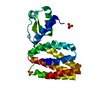
| ||||||||
|---|---|---|---|---|---|---|---|---|---|
| 1 | 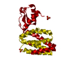
| ||||||||
| Unit cell |
|
-Components
| #1: Protein | Mass: 25027.408 Da / Num. of mol.: 1 Source method: isolated from a genetically manipulated source Source: (gene. exp.) Pseudomonas aeruginosa PAO1 (bacteria) / Gene: PA0797 / Production host: | ||||
|---|---|---|---|---|---|
| #2: Chemical | ChemComp-ZN /   Mass: 65.409 Da / Num. of mol.: 1 / Source method: obtained synthetically / Formula: Zn / Feature type: SUBJECT OF INVESTIGATION Mass: 65.409 Da / Num. of mol.: 1 / Source method: obtained synthetically / Formula: Zn / Feature type: SUBJECT OF INVESTIGATION | ||||
| #3: Chemical |   Mass: 96.063 Da / Num. of mol.: 3 / Source method: obtained synthetically / Formula: SO4 / Feature type: SUBJECT OF INVESTIGATION Mass: 96.063 Da / Num. of mol.: 3 / Source method: obtained synthetically / Formula: SO4 / Feature type: SUBJECT OF INVESTIGATION#4: Water | ChemComp-HOH / |  Mass: 18.015 Da / Num. of mol.: 2 / Source method: isolated from a natural source / Formula: H2O Mass: 18.015 Da / Num. of mol.: 2 / Source method: isolated from a natural source / Formula: H2OHas ligand of interest | Y | |
-Experimental details
-Experiment
| Experiment | Method: X-RAY DIFFRACTION / Number of used crystals: 1 |
|---|
- Sample preparation
| Crystal | Density Matthews: 2.66 Å3/Da / Density % sol: 54 % / Description: block |
|---|---|
| Crystal grow | Temperature: 290 K / Method: vapor diffusion, hanging drop / Details: MES/Sodium hydroxide, Magnesium sulfate |
-Data collection
| Diffraction | Mean temperature: 100 K / Serial crystal experiment: N | |||||||||||||||||||||||||||||||||||||||||||||||||||||||||||||||||||||||||||||||||||||||||||||||||||
|---|---|---|---|---|---|---|---|---|---|---|---|---|---|---|---|---|---|---|---|---|---|---|---|---|---|---|---|---|---|---|---|---|---|---|---|---|---|---|---|---|---|---|---|---|---|---|---|---|---|---|---|---|---|---|---|---|---|---|---|---|---|---|---|---|---|---|---|---|---|---|---|---|---|---|---|---|---|---|---|---|---|---|---|---|---|---|---|---|---|---|---|---|---|---|---|---|---|---|---|---|
| Diffraction source | Source: SYNCHROTRON / Site: SSRF / Beamline: BL19U1 / Wavelength: 0.9793 Å | |||||||||||||||||||||||||||||||||||||||||||||||||||||||||||||||||||||||||||||||||||||||||||||||||||
| Detector | Type: MARMOSAIC 325 mm CCD / Detector: CCD / Date: Oct 18, 2021 | |||||||||||||||||||||||||||||||||||||||||||||||||||||||||||||||||||||||||||||||||||||||||||||||||||
| Radiation | Protocol: SINGLE WAVELENGTH / Monochromatic (M) / Laue (L): M / Scattering type: x-ray | |||||||||||||||||||||||||||||||||||||||||||||||||||||||||||||||||||||||||||||||||||||||||||||||||||
| Radiation wavelength | Wavelength: 0.9793 Å / Relative weight: 1 | |||||||||||||||||||||||||||||||||||||||||||||||||||||||||||||||||||||||||||||||||||||||||||||||||||
| Reflection | Resolution: 2.6→30 Å / Num. obs: 8283 / % possible obs: 98.6 % / Redundancy: 8.7 % / Biso Wilson estimate: 35.53 Å2 / Rmerge(I) obs: 0.115 / Rpim(I) all: 0.037 / Rrim(I) all: 0.121 / Χ2: 0.941 / Net I/σ(I): 15.1 / Num. measured all: 71992 | |||||||||||||||||||||||||||||||||||||||||||||||||||||||||||||||||||||||||||||||||||||||||||||||||||
| Reflection shell | Diffraction-ID: 1
|
- Processing
| Software |
| ||||||||||||||||||||||||||||||||||||||||
|---|---|---|---|---|---|---|---|---|---|---|---|---|---|---|---|---|---|---|---|---|---|---|---|---|---|---|---|---|---|---|---|---|---|---|---|---|---|---|---|---|---|
| Refinement | Method to determine structure: MOLECULAR REPLACEMENT Starting model: model generated by Alphafold2 Resolution: 2.61→29.38 Å / SU ML: 0.29 / Cross valid method: THROUGHOUT / σ(F): 1.49 / Phase error: 25.54 / Stereochemistry target values: ML
| ||||||||||||||||||||||||||||||||||||||||
| Solvent computation | Shrinkage radii: 0.9 Å / VDW probe radii: 1.11 Å / Solvent model: FLAT BULK SOLVENT MODEL | ||||||||||||||||||||||||||||||||||||||||
| Displacement parameters | Biso max: 132.47 Å2 / Biso mean: 49.669 Å2 / Biso min: 13.8 Å2 | ||||||||||||||||||||||||||||||||||||||||
| Refinement step | Cycle: final / Resolution: 2.61→29.38 Å
| ||||||||||||||||||||||||||||||||||||||||
| LS refinement shell | Refine-ID: X-RAY DIFFRACTION / Rfactor Rfree error: 0 / Total num. of bins used: 3
| ||||||||||||||||||||||||||||||||||||||||
| Refinement TLS params. | Method: refined / Origin x: -32.8019 Å / Origin y: -26.1226 Å / Origin z: -23.2835 Å
| ||||||||||||||||||||||||||||||||||||||||
| Refinement TLS group |
|
