 Movie
Movie Controller

[English] 日本語
 Yorodumi
- PDB-7xjc: Crystal structure of bacteriorhodopsin in the ground and K states... -
+ Open data

- Basic information
| Entry | Database: PDB / ID: 7xjc | ||||||
|---|---|---|---|---|---|---|---|
| Title | Crystal structure of bacteriorhodopsin in the ground and K states after green laser irradiation | ||||||
 Components Components | Bacteriorhodopsin | ||||||
 Keywords Keywords | PROTON TRANSPORT / proton pump / membrane protein | ||||||
| Function / homology |  Function and homology information Function and homology informationlight-driven active monoatomic ion transmembrane transporter activity / photoreceptor activity / phototransduction / monoatomic ion channel activity / proton transmembrane transport / plasma membrane Similarity search - Function | ||||||
| Biological species |  Halobacterium salinarum NRC-1 (Halophile) Halobacterium salinarum NRC-1 (Halophile) | ||||||
| Method |  X-RAY DIFFRACTION / SYNCHROTRON / MOLECULAR REPLACEMENT / Resolution: 1.33 Å X-RAY DIFFRACTION / SYNCHROTRON / MOLECULAR REPLACEMENT / Resolution: 1.33 Å | ||||||
 Authors Authors | Taguchi, S. / Niwa, S. / Takeda, K. | ||||||
| Funding support |  Japan, 1items Japan, 1items
| ||||||
 Citation Citation | Journal: Commun Biol / Year: 2023 Title: Detailed analysis of distorted retinal and its interaction with surrounding residues in the K intermediate of bacteriorhodopsin Authors: Taguchi, S. / Niwa, S. / Dao, H.A. / Tanaka, Y. / Takeda, R. / Fukai, S. / Hasegawa, K. / Takeda, K. | ||||||
| History |
|
- Structure visualization
| Structure viewer | Molecule: MolmilJmol/JSmol |
|---|
- Downloads & links
-Download
| PDBx/mmCIF format | 7xjc.cif.gz | 161.3 KB | Display | PDBx/mmCIF format |
|---|---|---|---|---|
| PDB format | pdb7xjc.ent.gz | 132.1 KB | Display | PDB format |
| PDBx/mmJSON format | 7xjc.json.gz | Tree view | PDBx/mmJSON format | |
| Others |  Other downloads Other downloads |
-Validation report
| Arichive directory | https://data.pdbj.org/pub/pdb/validation_reports/xj/7xjcftp://data.pdbj.org/pub/pdb/validation_reports/xj/7xjc | HTTPS FTP |
|---|
-Related structure data
| Related structure data |  7xjeC  5zilS C: citing same article ( S: Starting model for refinement |
|---|---|
| Similar structure data | |
| Experimental dataset #1 | Data reference: 10.51093/xrd-00332 / Data set type: diffraction image data |

-Links
 PDBj
PDBj


- Assembly
| Deposited unit | 
| ||||||||
|---|---|---|---|---|---|---|---|---|---|
| 1 | 
| ||||||||
| Unit cell |
|
-Components
| #1: Protein | Mass: 25190.729 Da / Num. of mol.: 1 / Source method: isolated from a natural source / Source: (natural) Halobacterium salinarum NRC-1 (Halophile) / Strain: ATCC 700922 / JCM 11081 / NRC-1 / References: UniProt: P02945 | ||||||||
|---|---|---|---|---|---|---|---|---|---|
| #2: Chemical | ChemComp-RET /   Mass: 284.436 Da / Num. of mol.: 1 / Source method: obtained synthetically / Formula: C20H28O Mass: 284.436 Da / Num. of mol.: 1 / Source method: obtained synthetically / Formula: C20H28O | ||||||||
| #3: Chemical | ChemComp-L2P / 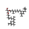  Mass: 653.157 Da / Num. of mol.: 14 / Source method: obtained synthetically / Formula: C43H88O3 Mass: 653.157 Da / Num. of mol.: 14 / Source method: obtained synthetically / Formula: C43H88O3#4: Chemical | ChemComp-SQU / 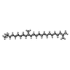  Mass: 380.734 Da / Num. of mol.: 4 / Source method: obtained synthetically / Formula: C27H56 Mass: 380.734 Da / Num. of mol.: 4 / Source method: obtained synthetically / Formula: C27H56#5: Water | ChemComp-HOH / |  Mass: 18.015 Da / Num. of mol.: 58 / Source method: isolated from a natural source / Formula: H2O Mass: 18.015 Da / Num. of mol.: 58 / Source method: isolated from a natural source / Formula: H2OHas ligand of interest | Y | Has protein modification | Y | |
-Experimental details
-Experiment
| Experiment | Method: X-RAY DIFFRACTION / Number of used crystals: 1 |
|---|
- Sample preparation
| Crystal | Density Matthews: 2.35 Å3/Da / Density % sol: 47.74 % |
|---|---|
| Crystal grow | Temperature: 293 K / Method: lipidic cubic phase / pH: 5.6 / Details: MO, 2.0-2.5 M Na/K phosphate pH 5.6 |
-Data collection
| Diffraction | Mean temperature: 15 K / Serial crystal experiment: N | ||||||||||||||||||||||||||||||||||||||||||||||||||||||||||||||||||||||||||||||||
|---|---|---|---|---|---|---|---|---|---|---|---|---|---|---|---|---|---|---|---|---|---|---|---|---|---|---|---|---|---|---|---|---|---|---|---|---|---|---|---|---|---|---|---|---|---|---|---|---|---|---|---|---|---|---|---|---|---|---|---|---|---|---|---|---|---|---|---|---|---|---|---|---|---|---|---|---|---|---|---|---|---|
| Diffraction source | Source: SYNCHROTRON / Site: SPring-8 / Beamline: BL41XU / Wavelength: 0.8 Å | ||||||||||||||||||||||||||||||||||||||||||||||||||||||||||||||||||||||||||||||||
| Detector | Type: DECTRIS EIGER X 16M / Detector: PIXEL / Date: Oct 6, 2020 | ||||||||||||||||||||||||||||||||||||||||||||||||||||||||||||||||||||||||||||||||
| Radiation | Protocol: SINGLE WAVELENGTH / Monochromatic (M) / Laue (L): M / Scattering type: x-ray | ||||||||||||||||||||||||||||||||||||||||||||||||||||||||||||||||||||||||||||||||
| Radiation wavelength | Wavelength: 0.8 Å / Relative weight: 1 | ||||||||||||||||||||||||||||||||||||||||||||||||||||||||||||||||||||||||||||||||
| Reflection | Resolution: 1.33→50 Å / Num. obs: 52175 / % possible obs: 99.9 % / Redundancy: 5.81 % / Biso Wilson estimate: 23.976 Å2 / CC1/2: 0.998 / Rmerge(I) obs: 0.074 / Rrim(I) all: 0.082 / Χ2: 0.823 / Net I/σ(I): 11.1 | ||||||||||||||||||||||||||||||||||||||||||||||||||||||||||||||||||||||||||||||||
| Reflection shell | Diffraction-ID: 1
|
- Processing
| Software |
| ||||||||||||||||||||
|---|---|---|---|---|---|---|---|---|---|---|---|---|---|---|---|---|---|---|---|---|---|
| Refinement | Method to determine structure: MOLECULAR REPLACEMENT Starting model: 5zil Resolution: 1.33→50 Å / Cross valid method: FREE R-VALUE
| ||||||||||||||||||||
| Displacement parameters | Biso max: 127.8 Å2 / Biso mean: 32.3397 Å2 / Biso min: 11.64 Å2 | ||||||||||||||||||||
| Refinement step | Cycle: LAST / Resolution: 1.33→50 Å
|
