- PDB-7xhx: Crystal structure of metallo-beta-lactamase IMP-6 -
+
Open data
ID or keywords:
Loading...
-
Basic information
Entry
Database: PDB / ID: 7xhx
Title
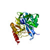
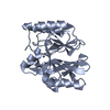
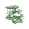
Crystal structure of metallo-beta-lactamase IMP-6
Components
Beta-lactamase
Keywords
HYDROLASE / Metallo-beta-lactamase / Carbapenemase / Zinc protein / Antimicrobial protein
Function / homology
Function and homology information
antibiotic catabolic process / beta-lactamase activity / beta-lactamase / periplasmic space / response to antibiotic / zinc ion binding Similarity search - Function
Beta-lactamases class B signature 2. / Beta-lactamases class B signature 1. / Beta-lactamase, class-B, conserved site / : / : / Metallo-beta-lactamase superfamily / Ribonuclease Z/Hydroxyacylglutathione hydrolase-like / Metallo-beta-lactamase; Chain A / Metallo-beta-lactamase superfamily / Metallo-beta-lactamase ...Beta-lactamases class B signature 2. / Beta-lactamases class B signature 1. / Beta-lactamase, class-B, conserved site / : / : / Metallo-beta-lactamase superfamily / Ribonuclease Z/Hydroxyacylglutathione hydrolase-like / Metallo-beta-lactamase; Chain A / Metallo-beta-lactamase superfamily / Metallo-beta-lactamase / Ribonuclease Z/Hydroxyacylglutathione hydrolase-like / 4-Layer Sandwich / Alpha Beta Similarity search - Domain/homology
Resolution: 1.7→48.3 Å / Cor.coef. Fo:Fc: 0.962 / Cor.coef. Fo:Fc free: 0.951 / SU B: 2.534 / SU ML: 0.082 / Cross valid method: THROUGHOUT / ESU R: 0.107 / ESU R Free: 0.106 / Stereochemistry target values: MAXIMUM LIKELIHOOD / Details: HYDROGENS HAVE BEEN ADDED IN THE RIDING POSITIONS
Rfactor
Num. reflection
% reflection
Selection details
Rfree
0.23782
5610
5 %
RANDOM
Rwork
0.20449
-
-
-
obs
0.20616
106591
99.95 %
-
Solvent computation
Ion probe radii: 0.8 Å / Shrinkage radii: 0.8 Å / VDW probe radii: 1.2 Å / Solvent model: MASK
 Movie
Movie Controller


 Open data
 Basic information
 Components
 Keywords
 Function and homology information
 X-RAY DIFFRACTION /
  Authors
 Japan, 1items
  Citation
 Structure visualization
 Downloads & links
 Other downloads
 PDBj
PDBj

 Assembly
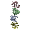

 Mass: 65.409 Da / Num. of mol.: 8 / Source method: obtained synthetically / Formula: Zn / Feature type: SUBJECT OF INVESTIGATION
 Mass: 18.015 Da / Num. of mol.: 377 / Source method: isolated from a natural source / Formula: H2O
 Sample preparation
 Processing