 Movie
Movie Controller

[English] 日本語
 Yorodumi
- PDB-7xhg: Crystal structure of the NTF2L domain of human G3BP1 in complex w... -
+ Open data

- Basic information
| Entry | Database: PDB / ID: 7xhg | ||||||
|---|---|---|---|---|---|---|---|
| Title | Crystal structure of the NTF2L domain of human G3BP1 in complex with the Caprin-1 derived peptide | ||||||
 Components Components |
| ||||||
 Keywords Keywords | RNA BINDING PROTEIN / Complex / Stress granules / Lipid-lipid phase separation | ||||||
| Function / homology |  Function and homology information Function and homology informationDNA/RNA helicase activity / ribosomal small subunit binding / positive regulation of type I interferon production / DNA helicase activity / stress granule assembly / molecular condensate scaffold activity / negative regulation of canonical Wnt signaling pathway / cytoplasmic stress granule / endonuclease activity / perikaryon ...DNA/RNA helicase activity / ribosomal small subunit binding / positive regulation of type I interferon production / DNA helicase activity / stress granule assembly / molecular condensate scaffold activity / negative regulation of canonical Wnt signaling pathway / cytoplasmic stress granule / endonuclease activity / perikaryon / defense response to virus / DNA helicase / Ras protein signal transduction / RNA helicase activity / RNA helicase / innate immune response / focal adhesion / mRNA binding / SARS-CoV-2 activates/modulates innate and adaptive immune responses / ATP hydrolysis activity / DNA binding / RNA binding / ATP binding / nucleus / cytosol / cytoplasm Similarity search - Function | ||||||
| Biological species |  Homo sapiens (human) Homo sapiens (human) | ||||||
| Method |  X-RAY DIFFRACTION / SYNCHROTRON / MOLECULAR REPLACEMENT / Resolution: 2.46 Å X-RAY DIFFRACTION / SYNCHROTRON / MOLECULAR REPLACEMENT / Resolution: 2.46 Å | ||||||
 Authors Authors | Dan, S. / Weimin, G. | ||||||
| Funding support |  China, 1items China, 1items
| ||||||
 Citation Citation | Journal: Proc.Natl.Acad.Sci.USA / Year: 2022 Title: Yin and yang regulation of stress granules by Caprin-1. Authors: Song, D. / Kuang, L. / Yang, L. / Wang, L. / Li, H. / Li, X. / Zhu, Z. / Shi, C. / Zhu, H. / Gong, W. | ||||||
| History |
|
- Structure visualization
| Structure viewer | Molecule: MolmilJmol/JSmol |
|---|
- Downloads & links
-Download
| PDBx/mmCIF format | 7xhg.cif.gz | 121.5 KB | Display | PDBx/mmCIF format |
|---|---|---|---|---|
| PDB format | pdb7xhg.ent.gz | 95.1 KB | Display | PDB format |
| PDBx/mmJSON format | 7xhg.json.gz | Tree view | PDBx/mmJSON format | |
| Others |  Other downloads Other downloads |
-Validation report
| Summary document | 7xhg_validation.pdf.gz | 473.3 KB | Display | wwPDB validaton report |
|---|---|---|---|---|
| Full document | 7xhg_full_validation.pdf.gz | 479.5 KB | Display | |
| Data in XML | 7xhg_validation.xml.gz | 21.5 KB | Display | |
| Data in CIF | 7xhg_validation.cif.gz | 29 KB | Display | |
| Arichive directory | https://data.pdbj.org/pub/pdb/validation_reports/xh/7xhgftp://data.pdbj.org/pub/pdb/validation_reports/xh/7xhg | HTTPS FTP |
-Related structure data
| Related structure data |  7xhfC  4fcjS S: Starting model for refinement C: citing same article ( |
|---|---|
| Similar structure data |
-Links
 PDBj
PDBj
- Assembly
| Deposited unit | 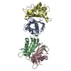
| ||||||||
|---|---|---|---|---|---|---|---|---|---|
| 1 | 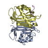
| ||||||||
| 2 | 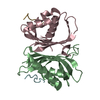
| ||||||||
| Unit cell |
|
-Components
| #1: Protein | Mass: 15899.076 Da / Num. of mol.: 4 Source method: isolated from a genetically manipulated source Source: (gene. exp.) Homo sapiens (human) / Gene: G3BP1, G3BP / Production host:  #2: Protein/peptide | Mass: 1227.386 Da / Num. of mol.: 3 Source method: isolated from a genetically manipulated source Source: (gene. exp.) Homo sapiens (human) / Production host: #3: Water | ChemComp-HOH / |  Mass: 18.015 Da / Num. of mol.: 41 / Source method: isolated from a natural source / Formula: H2O Mass: 18.015 Da / Num. of mol.: 41 / Source method: isolated from a natural source / Formula: H2O |
|---|
-Experimental details
-Experiment
| Experiment | Method: X-RAY DIFFRACTION / Number of used crystals: 1 |
|---|
- Sample preparation
| Crystal | Density Matthews: 2.34 Å3/Da / Density % sol: 47.36 % |
|---|---|
| Crystal grow | Temperature: 289.15 K / Method: vapor diffusion, sitting drop Details: 0.2 M Sodium acetate trihydrate, 0.1 M Sodium cacodylate trihydrate pH 6.5, 30 % w/v Polyethylene glycol 8,000 |
-Data collection
| Diffraction | Mean temperature: 100 K / Serial crystal experiment: N |
|---|---|
| Diffraction source | Source: SYNCHROTRON / Site: SSRF / Beamline: BL17U1 / Wavelength: 0.97915 Å |
| Detector | Type: DECTRIS EIGER X 16M / Detector: PIXEL / Date: Oct 23, 2019 |
| Radiation | Protocol: SINGLE WAVELENGTH / Monochromatic (M) / Laue (L): M / Scattering type: x-ray |
| Radiation wavelength | Wavelength: 0.97915 Å / Relative weight: 1 |
| Reflection | Resolution: 2.45→85.88 Å / Num. obs: 21506 / % possible obs: 99.6 % / Redundancy: 3.4 % / CC1/2: 0.971 / Rmerge(I) obs: 0.106 / Rpim(I) all: 0.068 / Rrim(I) all: 0.126 / Net I/σ(I): 28.2 |
| Reflection shell | Resolution: 2.45→2.49 Å / Rmerge(I) obs: 0.532 / Mean I/σ(I) obs: 4.1 / Num. unique obs: 1065 / CC1/2: 0.818 / Rpim(I) all: 0.333 / Rrim(I) all: 0.629 |
- Processing
| Software |
| ||||||||||||||||||||||||||||||||||||||||||||||||||||||||||||
|---|---|---|---|---|---|---|---|---|---|---|---|---|---|---|---|---|---|---|---|---|---|---|---|---|---|---|---|---|---|---|---|---|---|---|---|---|---|---|---|---|---|---|---|---|---|---|---|---|---|---|---|---|---|---|---|---|---|---|---|---|---|
| Refinement | Method to determine structure: MOLECULAR REPLACEMENT Starting model: 4fcj Resolution: 2.46→85.88 Å / Cor.coef. Fo:Fc: 0.937 / Cor.coef. Fo:Fc free: 0.882 / SU B: 12.939 / SU ML: 0.284 / Cross valid method: THROUGHOUT / σ(F): 0 / ESU R: 0.706 / ESU R Free: 0.323 / Stereochemistry target values: MAXIMUM LIKELIHOOD Details: HYDROGENS HAVE BEEN ADDED IN THE RIDING POSITIONS U VALUES : REFINED INDIVIDUALLY
| ||||||||||||||||||||||||||||||||||||||||||||||||||||||||||||
| Solvent computation | Ion probe radii: 0.8 Å / Shrinkage radii: 0.8 Å / VDW probe radii: 1.2 Å / Solvent model: MASK | ||||||||||||||||||||||||||||||||||||||||||||||||||||||||||||
| Displacement parameters | Biso max: 90.24 Å2 / Biso mean: 39.622 Å2 / Biso min: 16.93 Å2
| ||||||||||||||||||||||||||||||||||||||||||||||||||||||||||||
| Refinement step | Cycle: final / Resolution: 2.46→85.88 Å
| ||||||||||||||||||||||||||||||||||||||||||||||||||||||||||||
| Refine LS restraints |
| ||||||||||||||||||||||||||||||||||||||||||||||||||||||||||||
| LS refinement shell | Resolution: 2.46→2.519 Å / Rfactor Rfree error: 0
|
