 Movie
Movie Controller

[English] 日本語
 Yorodumi
- PDB-7xh4: Dihydrofolate Reductase-like Protein SacH in safracin biosynthesi... -
+ Open data

- Basic information
| Entry | Database: PDB / ID: 7xh4 | ||||||
|---|---|---|---|---|---|---|---|
| Title | Dihydrofolate Reductase-like Protein SacH in safracin biosynthesis complex with safracin A | ||||||
 Components Components | Uncharacterized protein sfcH | ||||||
 Keywords Keywords | OXIDOREDUCTASE / reductase / safracin biosynthesis / self-resistance / complex structure. | ||||||
| Function / homology |  Function and homology information Function and homology information5-amino-6-(5-phosphoribosylamino)uracil reductase activity / riboflavin biosynthetic process / nucleotide binding Similarity search - Function | ||||||
| Biological species |  Pseudomonas fluorescens (bacteria) Pseudomonas fluorescens (bacteria) | ||||||
| Method |  X-RAY DIFFRACTION / MOLECULAR REPLACEMENT / Resolution: 2.2 Å X-RAY DIFFRACTION / MOLECULAR REPLACEMENT / Resolution: 2.2 Å | ||||||
 Authors Authors | Ma, X. / Shao, N. / Zhang, Y. / Yang, D. / Ma, M. / Tang, G. | ||||||
| Funding support |  China, 1items China, 1items
| ||||||
 Citation Citation | Journal: Acta Pharm Sin B / Year: 2023 Title: Dihydrofolate reductase-like protein inactivates hemiaminal pharmacophore for self-resistance in safracin biosynthesis. Authors: Shao, N. / Ma, X. / Zhang, Y.Y. / Yang, D. / Ma, M. / Tang, G.L. | ||||||
| History |
|
- Structure visualization
| Structure viewer | Molecule: MolmilJmol/JSmol |
|---|
- Downloads & links
-Download
| PDBx/mmCIF format | 7xh4.cif.gz | 55.5 KB | Display | PDBx/mmCIF format |
|---|---|---|---|---|
| PDB format | pdb7xh4.ent.gz | 35.8 KB | Display | PDB format |
| PDBx/mmJSON format | 7xh4.json.gz | Tree view | PDBx/mmJSON format | |
| Others |  Other downloads Other downloads |
-Validation report
| Arichive directory | https://data.pdbj.org/pub/pdb/validation_reports/xh/7xh4ftp://data.pdbj.org/pub/pdb/validation_reports/xh/7xh4 | HTTPS FTP |
|---|
-Related structure data
| Related structure data | 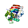 7xh2C 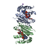 2xw7S S: Starting model for refinement C: citing same article ( |
|---|---|
| Similar structure data |
-Links
 PDBj
PDBj

- Assembly
| Deposited unit | 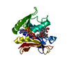
| ||||||||
|---|---|---|---|---|---|---|---|---|---|
| 1 |
| ||||||||
| Unit cell |
|
-Components
| #1: Protein | Mass: 22018.842 Da / Num. of mol.: 1 Source method: isolated from a genetically manipulated source Source: (gene. exp.) Pseudomonas fluorescens (bacteria)Production host: References: UniProt: Q5JCL3 |
|---|---|
| #2: Chemical | ChemComp-EN7 / 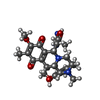  Mass: 524.609 Da / Num. of mol.: 1 / Source method: obtained synthetically / Formula: C28H36N4O6 / Feature type: SUBJECT OF INVESTIGATION Mass: 524.609 Da / Num. of mol.: 1 / Source method: obtained synthetically / Formula: C28H36N4O6 / Feature type: SUBJECT OF INVESTIGATION |
| #3: Chemical | ChemComp-NDP /   Mass: 745.421 Da / Num. of mol.: 1 / Source method: obtained synthetically / Formula: C21H30N7O17P3 / Feature type: SUBJECT OF INVESTIGATION Mass: 745.421 Da / Num. of mol.: 1 / Source method: obtained synthetically / Formula: C21H30N7O17P3 / Feature type: SUBJECT OF INVESTIGATION |
| #4: Water | ChemComp-HOH /  Mass: 18.015 Da / Num. of mol.: 69 / Source method: isolated from a natural source / Formula: H2O Mass: 18.015 Da / Num. of mol.: 69 / Source method: isolated from a natural source / Formula: H2O |
| Has ligand of interest | Y |
-Experimental details
-Experiment
| Experiment | Method: X-RAY DIFFRACTION / Number of used crystals: 1 |
|---|
- Sample preparation
| Crystal | Density Matthews: 2.44 Å3/Da / Density % sol: 49.49 % |
|---|---|
| Crystal grow | Temperature: 289 K / Method: vapor diffusion, hanging drop / Details: 28% (w/v) PEG 1500, 100mM PCB buffer pH 7.0 |
-Data collection
| Diffraction | Mean temperature: 100 K / Serial crystal experiment: N |
|---|---|
| Diffraction source | Source: ROTATING ANODE / Type: RIGAKU PhotonJet-R / Wavelength: 1.54184 Å |
| Detector | Type: RIGAKU HyPix-6000HE / Detector: PIXEL / Date: Sep 2, 2021 |
| Radiation | Protocol: SINGLE WAVELENGTH / Monochromatic (M) / Laue (L): M / Scattering type: x-ray |
| Radiation wavelength | Wavelength: 1.54184 Å / Relative weight: 1 |
| Reflection | Resolution: 2.2→29.76 Å / Num. obs: 11851 / % possible obs: 99.7 % / Redundancy: 20 % / Rmerge(I) obs: 0.078 / Net I/σ(I): 55.28 |
| Reflection shell | Resolution: 2.2→2.28 Å / Rmerge(I) obs: 0.496 / Num. unique obs: 1186 |
- Processing
| Software |
| |||||||||||||||||||||||||||||||||||||||||||||||||||||||||||||||
|---|---|---|---|---|---|---|---|---|---|---|---|---|---|---|---|---|---|---|---|---|---|---|---|---|---|---|---|---|---|---|---|---|---|---|---|---|---|---|---|---|---|---|---|---|---|---|---|---|---|---|---|---|---|---|---|---|---|---|---|---|---|---|---|---|
| Refinement | Method to determine structure: MOLECULAR REPLACEMENT Starting model: 2XW7 Resolution: 2.2→29.76 Å / SU ML: 0.34 / Cross valid method: THROUGHOUT / σ(F): 1.34 / Phase error: 28.32 / Stereochemistry target values: ML
| |||||||||||||||||||||||||||||||||||||||||||||||||||||||||||||||
| Solvent computation | Shrinkage radii: 0.9 Å / VDW probe radii: 1.11 Å / Solvent model: FLAT BULK SOLVENT MODEL | |||||||||||||||||||||||||||||||||||||||||||||||||||||||||||||||
| Displacement parameters | Biso max: 74.84 Å2 / Biso mean: 34.9333 Å2 / Biso min: 16.02 Å2 | |||||||||||||||||||||||||||||||||||||||||||||||||||||||||||||||
| Refinement step | Cycle: final / Resolution: 2.2→29.76 Å
| |||||||||||||||||||||||||||||||||||||||||||||||||||||||||||||||
| LS refinement shell | Refine-ID: X-RAY DIFFRACTION / Rfactor Rfree error: 0 / Total num. of bins used: 8
|
