 Movie
Movie Controller

[English] 日本語
 Yorodumi
- PDB-7xgn: Quinolinate Phosphoribosyl Transferase (QAPRTase) from Streptomyc... -
+ Open data

- Basic information
| Entry | Database: PDB / ID: 7xgn | ||||||
|---|---|---|---|---|---|---|---|
| Title | Quinolinate Phosphoribosyl Transferase (QAPRTase) from Streptomyces pyridomyceticus NRRL B-2517 in complex with Nicotinic Acid (NA) | ||||||
 Components Components | Quinolinate Phosphoribosyl Transferase | ||||||
 Keywords Keywords | TRANSFERASE / nicotinamide adenine dinucleotide / pyridomycin / quinolinic acid | ||||||
| Function / homology | Aldolase class I / TIM Barrel / Alpha-Beta Barrel / Alpha Beta / NICOTINIC ACID Function and homology information Function and homology information | ||||||
| Biological species |  Streptomyces pyridomyceticus (bacteria) Streptomyces pyridomyceticus (bacteria) | ||||||
| Method |  X-RAY DIFFRACTION / SYNCHROTRON / MOLECULAR REPLACEMENT / Resolution: 2.6 Å X-RAY DIFFRACTION / SYNCHROTRON / MOLECULAR REPLACEMENT / Resolution: 2.6 Å | ||||||
 Authors Authors | Zhou, Z. / Yang, X. / Huang, T. / Wang, X. / Liang, R. / Zheng, J. / Dai, S. / Lin, S. / Deng, Z. | ||||||
| Funding support |  China, 1items China, 1items
| ||||||
 Citation Citation | Journal: Acs Chem.Biol. / Year: 2023 Title: Bifunctional NadC Homologue PyrZ Catalyzes Nicotinic Acid Formation in Pyridomycin Biosynthesis. Authors: Zhou, Z. / Yang, X. / Huang, T. / Zheng, J. / Deng, Z. / Dai, S. / Lin, S. | ||||||
| History |
|
- Structure visualization
| Structure viewer | Molecule: MolmilJmol/JSmol |
|---|
- Downloads & links
-Download
| PDBx/mmCIF format | 7xgn.cif.gz | 145.3 KB | Display | PDBx/mmCIF format |
|---|---|---|---|---|
| PDB format | pdb7xgn.ent.gz | 92 KB | Display | PDB format |
| PDBx/mmJSON format | 7xgn.json.gz | Tree view | PDBx/mmJSON format | |
| Others |  Other downloads Other downloads |
-Validation report
| Summary document | 7xgn_validation.pdf.gz | 465.8 KB | Display | wwPDB validaton report |
|---|---|---|---|---|
| Full document | 7xgn_full_validation.pdf.gz | 470.5 KB | Display | |
| Data in XML | 7xgn_validation.xml.gz | 22 KB | Display | |
| Data in CIF | 7xgn_validation.cif.gz | 30.3 KB | Display | |
| Arichive directory | https://data.pdbj.org/pub/pdb/validation_reports/xg/7xgnftp://data.pdbj.org/pub/pdb/validation_reports/xg/7xgn | HTTPS FTP |
-Related structure data
| Related structure data |  7xglC  7xgmC 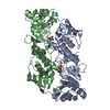 1qpoS S: Starting model for refinement C: citing same article ( |
|---|---|
| Similar structure data |
-Links
 PDBj
PDBj- Assembly
| Deposited unit | 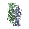
| ||||||||||||
|---|---|---|---|---|---|---|---|---|---|---|---|---|---|
| 1 |
| ||||||||||||
| Unit cell |
|
-Components
| #1: Protein | Mass: 32412.721 Da / Num. of mol.: 2 Source method: isolated from a genetically manipulated source Source: (gene. exp.) Streptomyces pyridomyceticus (bacteria)Production host: #2: Chemical |   Mass: 96.063 Da / Num. of mol.: 2 / Source method: obtained synthetically / Formula: SO4 Mass: 96.063 Da / Num. of mol.: 2 / Source method: obtained synthetically / Formula: SO4#3: Chemical | ChemComp-CL /   Mass: 35.453 Da / Num. of mol.: 5 / Source method: obtained synthetically / Formula: Cl Mass: 35.453 Da / Num. of mol.: 5 / Source method: obtained synthetically / Formula: Cl#4: Chemical |   Mass: 123.109 Da / Num. of mol.: 2 / Source method: obtained synthetically / Formula: C6H5NO2 Mass: 123.109 Da / Num. of mol.: 2 / Source method: obtained synthetically / Formula: C6H5NO2#5: Water | ChemComp-HOH / |  Mass: 18.015 Da / Num. of mol.: 49 / Source method: isolated from a natural source / Formula: H2O Mass: 18.015 Da / Num. of mol.: 49 / Source method: isolated from a natural source / Formula: H2OHas ligand of interest | N | |
|---|
-Experimental details
-Experiment
| Experiment | Method: X-RAY DIFFRACTION / Number of used crystals: 1 |
|---|
- Sample preparation
| Crystal | Density Matthews: 3.82 Å3/Da / Density % sol: 67.83 % |
|---|---|
| Crystal grow | Temperature: 293 K / Method: vapor diffusion, hanging drop / Details: 0.1M Citric acid, pH 5.0, 1.6M Ammonium sulfate |
-Data collection
| Diffraction | Mean temperature: 193 K / Serial crystal experiment: N |
|---|---|
| Diffraction source | Source: SYNCHROTRON / Site: SSRF / Beamline: BL18U1 / Wavelength: 0.97915 Å |
| Detector | Type: DECTRIS PILATUS3 6M / Detector: PIXEL / Date: Jun 18, 2020 |
| Radiation | Protocol: SINGLE WAVELENGTH / Monochromatic (M) / Laue (L): M / Scattering type: x-ray |
| Radiation wavelength | Wavelength: 0.97915 Å / Relative weight: 1 |
| Reflection | Resolution: 2.6→40.46 Å / Num. obs: 31172 / % possible obs: 96.3 % / Redundancy: 4.6 % / Biso Wilson estimate: 43.48 Å2 / CC1/2: 0.985 / Net I/σ(I): 12.8 |
| Reflection shell | Resolution: 2.6→2.7 Å / Num. unique obs: 1506 / CC1/2: 0.787 |
- Processing
| Software |
| |||||||||||||||||||||||||||||||||||||||||||||||||||||||||||||||||||||||||||||
|---|---|---|---|---|---|---|---|---|---|---|---|---|---|---|---|---|---|---|---|---|---|---|---|---|---|---|---|---|---|---|---|---|---|---|---|---|---|---|---|---|---|---|---|---|---|---|---|---|---|---|---|---|---|---|---|---|---|---|---|---|---|---|---|---|---|---|---|---|---|---|---|---|---|---|---|---|---|---|
| Refinement | Method to determine structure: MOLECULAR REPLACEMENT Starting model: 1QPO Resolution: 2.6→40.46 Å / SU ML: 0.3307 / Cross valid method: FREE R-VALUE / σ(F): 1.36 / Phase error: 22.8927 Stereochemistry target values: GeoStd + Monomer Library + CDL v1.2
| |||||||||||||||||||||||||||||||||||||||||||||||||||||||||||||||||||||||||||||
| Solvent computation | Shrinkage radii: 0.9 Å / VDW probe radii: 1.11 Å / Solvent model: FLAT BULK SOLVENT MODEL | |||||||||||||||||||||||||||||||||||||||||||||||||||||||||||||||||||||||||||||
| Displacement parameters | Biso mean: 40.77 Å2 | |||||||||||||||||||||||||||||||||||||||||||||||||||||||||||||||||||||||||||||
| Refinement step | Cycle: LAST / Resolution: 2.6→40.46 Å
| |||||||||||||||||||||||||||||||||||||||||||||||||||||||||||||||||||||||||||||
| Refine LS restraints |
| |||||||||||||||||||||||||||||||||||||||||||||||||||||||||||||||||||||||||||||
| LS refinement shell |
|
