 Movie
Movie Controller

[English] 日本語
 Yorodumi
- PDB-7xfw: Crystal structure of the ternary complex of Peptidoglycan recogni... -
+ Open data

- Basic information
| Entry | Database: PDB / ID: 7xfw | |||||||||
|---|---|---|---|---|---|---|---|---|---|---|
| Title | Crystal structure of the ternary complex of Peptidoglycan recognition protein, PGRP-S with hexanoic and tartaric acids at 2.07 A resolution. | |||||||||
 Components Components | Peptidoglycan recognition protein 1 | |||||||||
 Keywords Keywords | IMMUNE SYSTEM / CPGRP-S / PGRP-S | |||||||||
| Function / homology |  Function and homology information Function and homology informationpeptidoglycan immune receptor activity / N-acetylmuramoyl-L-alanine amidase activity / peptidoglycan binding / negative regulation of cytokine production / detection of bacterium / peptidoglycan catabolic process / defense response to Gram-positive bacterium / innate immune response / zinc ion binding / extracellular region Similarity search - Function | |||||||||
| Biological species |  | |||||||||
| Method |  X-RAY DIFFRACTION / SYNCHROTRON / MOLECULAR REPLACEMENT / Resolution: 2.07 Å X-RAY DIFFRACTION / SYNCHROTRON / MOLECULAR REPLACEMENT / Resolution: 2.07 Å | |||||||||
 Authors Authors | Maurya, A. / Singh, P.K. / Viswanathan, V. / Sharma, P. / Sharma, S. / Singh, T.P. | |||||||||
| Funding support | 1items
| |||||||||
 Citation Citation | Journal: Biochim Biophys Acta Proteins Proteom / Year: 2022 Title: Structure of the complex of camel peptidoglycan recognition protein-S with hexanoic acid reveals novel features of the versatile ligand-binding site at the dimeric interface. Authors: Maurya, A. / Sharma, P. / Singh, P.K. / Viswanathan, V. / Kaur, P. / Sharma, S. / Singh, T.P. | |||||||||
| History |
|
- Structure visualization
| Structure viewer | Molecule: MolmilJmol/JSmol |
|---|
- Downloads & links
-Download
| PDBx/mmCIF format | 7xfw.cif.gz | 156.5 KB | Display | PDBx/mmCIF format |
|---|---|---|---|---|
| PDB format | pdb7xfw.ent.gz | 119.6 KB | Display | PDB format |
| PDBx/mmJSON format | 7xfw.json.gz | Tree view | PDBx/mmJSON format | |
| Others |  Other downloads Other downloads |
-Validation report
| Summary document | 7xfw_validation.pdf.gz | 3.7 MB | Display | wwPDB validaton report |
|---|---|---|---|---|
| Full document | 7xfw_full_validation.pdf.gz | 3.9 MB | Display | |
| Data in XML | 7xfw_validation.xml.gz | 32.3 KB | Display | |
| Data in CIF | 7xfw_validation.cif.gz | 45.8 KB | Display | |
| Arichive directory | https://data.pdbj.org/pub/pdb/validation_reports/xf/7xfwftp://data.pdbj.org/pub/pdb/validation_reports/xf/7xfw | HTTPS FTP |
-Related structure data
| Related structure data |  5dwfS S: Starting model for refinement |
|---|---|
| Similar structure data |
-Links
 PDBj
PDBj
- Assembly
| Deposited unit | 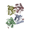
| ||||||||||||
|---|---|---|---|---|---|---|---|---|---|---|---|---|---|
| 1 | 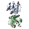
| ||||||||||||
| 2 | 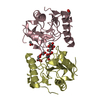
| ||||||||||||
| Unit cell |
| ||||||||||||
| Components on special symmetry positions |
|
-Components
-Protein , 1 types, 4 molecules ABCD
| #1: Protein | Mass: 19011.459 Da / Num. of mol.: 4 / Source method: isolated from a natural source / Source: (natural) |
|---|
-Non-polymers , 7 types, 482 molecules 


| #2: Chemical |  Mass: 150.087 Da / Num. of mol.: 3 / Source method: obtained synthetically / Formula: C4H6O6 / Feature type: SUBJECT OF INVESTIGATION Mass: 150.087 Da / Num. of mol.: 3 / Source method: obtained synthetically / Formula: C4H6O6 / Feature type: SUBJECT OF INVESTIGATION#3: Chemical | ChemComp-EDO /  Mass: 62.068 Da / Num. of mol.: 6 / Source method: obtained synthetically / Formula: C2H6O2 / Feature type: SUBJECT OF INVESTIGATION Mass: 62.068 Da / Num. of mol.: 6 / Source method: obtained synthetically / Formula: C2H6O2 / Feature type: SUBJECT OF INVESTIGATION#4: Chemical |  Mass: 59.044 Da / Num. of mol.: 3 / Source method: obtained synthetically / Formula: C2H3O2 / Feature type: SUBJECT OF INVESTIGATION Mass: 59.044 Da / Num. of mol.: 3 / Source method: obtained synthetically / Formula: C2H3O2 / Feature type: SUBJECT OF INVESTIGATION#5: Chemical | ChemComp-CL / |  Mass: 35.453 Da / Num. of mol.: 1 / Source method: obtained synthetically / Formula: Cl / Feature type: SUBJECT OF INVESTIGATION Mass: 35.453 Da / Num. of mol.: 1 / Source method: obtained synthetically / Formula: Cl / Feature type: SUBJECT OF INVESTIGATION#6: Chemical | ChemComp-6NA / |  Mass: 116.158 Da / Num. of mol.: 1 / Source method: obtained synthetically / Formula: C6H12O2 / Feature type: SUBJECT OF INVESTIGATION Mass: 116.158 Da / Num. of mol.: 1 / Source method: obtained synthetically / Formula: C6H12O2 / Feature type: SUBJECT OF INVESTIGATION#7: Chemical | ChemComp-GOL / |  Mass: 92.094 Da / Num. of mol.: 1 / Source method: obtained synthetically / Formula: C3H8O3 / Feature type: SUBJECT OF INVESTIGATION Mass: 92.094 Da / Num. of mol.: 1 / Source method: obtained synthetically / Formula: C3H8O3 / Feature type: SUBJECT OF INVESTIGATION#8: Water | ChemComp-HOH / | Mass: 18.015 Da / Num. of mol.: 467 / Source method: isolated from a natural source / Formula: H2O |
|---|
-Details
| Has ligand of interest | Y |
|---|
-Experimental details
-Experiment
| Experiment | Method: X-RAY DIFFRACTION / Number of used crystals: 1 |
|---|
- Sample preparation
| Crystal | Density Matthews: 2.39 Å3/Da / Density % sol: 48.51 % |
|---|---|
| Crystal grow | Temperature: 298 K / Method: vapor diffusion, hanging drop / pH: 8.5 Details: 10% PEG 3350 0.2M Sodium potassium tartrate 20% Glycerol |
-Data collection
| Diffraction | Mean temperature: 100 K / Serial crystal experiment: N |
|---|---|
| Diffraction source | Source: SYNCHROTRON / Site: ESRF  / Beamline: MASSIF-1 / Wavelength: 0.97 Å / Beamline: MASSIF-1 / Wavelength: 0.97 Å |
| Detector | Type: DECTRIS PILATUS3 2M / Detector: PIXEL / Date: Nov 7, 2018 |
| Radiation | Monochromator: M / Protocol: SINGLE WAVELENGTH / Monochromatic (M) / Laue (L): M / Scattering type: x-ray |
| Radiation wavelength | Wavelength: 0.97 Å / Relative weight: 1 |
| Reflection | Resolution: 2.07→46.36 Å / Num. obs: 45175 / % possible obs: 99.82 % / Redundancy: 6.94 % / CC1/2: 0.99 / Rpim(I) all: 0.041 / Net I/σ(I): 8.13 |
| Reflection shell | Resolution: 2.07→2.12 Å / Redundancy: 6 % / Mean I/σ(I) obs: 1.9 / Num. unique obs: 1297 / CC1/2: 0.869 / Rpim(I) all: 0.433 / % possible all: 99.79 |
- Processing
| Software |
| |||||||||||||||||||||||||||||||||||||||||||||||||||||||||||||||||||||||||||||||||||||||||||||||||||||||||||||||||||||||||||||||||||||||||||||||||||||||||||
|---|---|---|---|---|---|---|---|---|---|---|---|---|---|---|---|---|---|---|---|---|---|---|---|---|---|---|---|---|---|---|---|---|---|---|---|---|---|---|---|---|---|---|---|---|---|---|---|---|---|---|---|---|---|---|---|---|---|---|---|---|---|---|---|---|---|---|---|---|---|---|---|---|---|---|---|---|---|---|---|---|---|---|---|---|---|---|---|---|---|---|---|---|---|---|---|---|---|---|---|---|---|---|---|---|---|---|---|---|---|---|---|---|---|---|---|---|---|---|---|---|---|---|---|---|---|---|---|---|---|---|---|---|---|---|---|---|---|---|---|---|---|---|---|---|---|---|---|---|---|---|---|---|---|---|---|---|
| Refinement | Method to determine structure: MOLECULAR REPLACEMENT Starting model: 5DWF Resolution: 2.07→46.358 Å / Cor.coef. Fo:Fc: 0.958 / Cor.coef. Fo:Fc free: 0.928 / SU B: 7.577 / SU ML: 0.188 / Cross valid method: FREE R-VALUE / ESU R: 0.22 / ESU R Free: 0.195 Details: Hydrogens have been added in their riding positions
| |||||||||||||||||||||||||||||||||||||||||||||||||||||||||||||||||||||||||||||||||||||||||||||||||||||||||||||||||||||||||||||||||||||||||||||||||||||||||||
| Solvent computation | Ion probe radii: 0.8 Å / Shrinkage radii: 0.8 Å / VDW probe radii: 1.2 Å / Solvent model: MASK BULK SOLVENT | |||||||||||||||||||||||||||||||||||||||||||||||||||||||||||||||||||||||||||||||||||||||||||||||||||||||||||||||||||||||||||||||||||||||||||||||||||||||||||
| Displacement parameters | Biso mean: 36.53 Å2
| |||||||||||||||||||||||||||||||||||||||||||||||||||||||||||||||||||||||||||||||||||||||||||||||||||||||||||||||||||||||||||||||||||||||||||||||||||||||||||
| Refinement step | Cycle: LAST / Resolution: 2.07→46.358 Å
| |||||||||||||||||||||||||||||||||||||||||||||||||||||||||||||||||||||||||||||||||||||||||||||||||||||||||||||||||||||||||||||||||||||||||||||||||||||||||||
| Refine LS restraints |
| |||||||||||||||||||||||||||||||||||||||||||||||||||||||||||||||||||||||||||||||||||||||||||||||||||||||||||||||||||||||||||||||||||||||||||||||||||||||||||
| LS refinement shell |
|
