 Movie
Movie Controller

[English] 日本語
 Yorodumi
- PDB-7xef: Crystal Structure of the Y53F/N55A mutant of LEH complexed with (... -
+ Open data

- Basic information
| Entry | Database: PDB / ID: 7xef | ||||||
|---|---|---|---|---|---|---|---|
| Title | Crystal Structure of the Y53F/N55A mutant of LEH complexed with (R)-(1-benzyl-3-phenylpyrrolidin-3-yl)methanol | ||||||
 Components Components | Limonene-1,2-epoxide hydrolase | ||||||
 Keywords Keywords | HYDROLASE / limonene-1 / 2-epoxide hydrolase / mutant / Rhodococcus erythropolis | ||||||
| Function / homology | limonene-1,2-epoxide hydrolase / limonene-1,2-epoxide hydrolase activity / Limonene-1,2-epoxide hydrolase / Limonene-1,2-epoxide hydrolase catalytic domain / NTF2-like domain superfamily / Chem-D0I / DI(HYDROXYETHYL)ETHER / Limonene-1,2-epoxide hydrolase Function and homology information Function and homology information | ||||||
| Biological species |  Rhodococcus erythropolis (bacteria) Rhodococcus erythropolis (bacteria) | ||||||
| Method |  X-RAY DIFFRACTION / MOLECULAR REPLACEMENT / Resolution: 1.816 Å X-RAY DIFFRACTION / MOLECULAR REPLACEMENT / Resolution: 1.816 Å | ||||||
 Authors Authors | Qu, G. / Li, X. / Sun, Z.T. | ||||||
| Funding support | 1items
| ||||||
 Citation Citation | Journal: Nat Commun / Year: 2022 Title: Rational enzyme design for enabling biocatalytic Baldwin cyclization and asymmetric synthesis of chiral heterocycles. Authors: Li, J.K. / Qu, G. / Li, X. / Tian, Y. / Cui, C. / Zhang, F.G. / Zhang, W. / Ma, J.A. / Reetz, M.T. / Sun, Z. | ||||||
| History |
|
- Structure visualization
| Structure viewer | Molecule: MolmilJmol/JSmol |
|---|
- Downloads & links
-Download
| PDBx/mmCIF format | 7xef.cif.gz | 151.5 KB | Display | PDBx/mmCIF format |
|---|---|---|---|---|
| PDB format | pdb7xef.ent.gz | 116.1 KB | Display | PDB format |
| PDBx/mmJSON format | 7xef.json.gz | Tree view | PDBx/mmJSON format | |
| Others |  Other downloads Other downloads |
-Validation report
| Arichive directory | https://data.pdbj.org/pub/pdb/validation_reports/xe/7xefftp://data.pdbj.org/pub/pdb/validation_reports/xe/7xef | HTTPS FTP |
|---|
-Related structure data
| Related structure data |  7vwdC  7vwmC 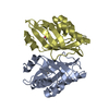 7vx2C  7xeeC  1nwwS S: Starting model for refinement C: citing same article ( |
|---|---|
| Similar structure data |
-Links
 PDBj
PDBj
- Assembly
| Deposited unit | 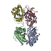
| ||||||||
|---|---|---|---|---|---|---|---|---|---|
| 1 | 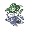
| ||||||||
| 2 | 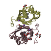
| ||||||||
| Unit cell |
|
-Components
-Protein , 1 types, 4 molecules ABCD
| #1: Protein | Mass: 17304.434 Da / Num. of mol.: 4 / Mutation: Y53F,N55A Source method: isolated from a genetically manipulated source Source: (gene. exp.) Rhodococcus erythropolis (bacteria) / Gene: limA / Production host: |
|---|
-Non-polymers , 5 types, 719 molecules 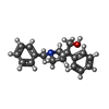


| #2: Chemical | ChemComp-D0I / [(  Mass: 267.365 Da / Num. of mol.: 4 / Source method: obtained synthetically / Formula: C18H21NO / Feature type: SUBJECT OF INVESTIGATION Mass: 267.365 Da / Num. of mol.: 4 / Source method: obtained synthetically / Formula: C18H21NO / Feature type: SUBJECT OF INVESTIGATION#3: Chemical | ChemComp-EDO /  Mass: 62.068 Da / Num. of mol.: 7 / Source method: obtained synthetically / Formula: C2H6O2 Mass: 62.068 Da / Num. of mol.: 7 / Source method: obtained synthetically / Formula: C2H6O2#4: Chemical | ChemComp-NA /  Mass: 22.990 Da / Num. of mol.: 6 / Source method: obtained synthetically / Formula: Na Mass: 22.990 Da / Num. of mol.: 6 / Source method: obtained synthetically / Formula: Na#5: Chemical | ChemComp-PEG / |  Mass: 106.120 Da / Num. of mol.: 1 / Source method: isolated from a natural source / Formula: C4H10O3 Mass: 106.120 Da / Num. of mol.: 1 / Source method: isolated from a natural source / Formula: C4H10O3#6: Water | ChemComp-HOH / | Mass: 18.015 Da / Num. of mol.: 701 / Source method: isolated from a natural source / Formula: H2O |
|---|
-Details
| Has ligand of interest | Y |
|---|
-Experimental details
-Experiment
| Experiment | Method: X-RAY DIFFRACTION / Number of used crystals: 1 |
|---|
- Sample preparation
| Crystal | Density Matthews: 2.01 Å3/Da / Density % sol: 38.92 % |
|---|---|
| Crystal grow | Temperature: 293 K / Method: vapor diffusion, sitting drop / pH: 7.5 / Details: NaCl, BIS-TRIS pH5.5, Polyethylene glycol 3350 |
-Data collection
| Diffraction | Mean temperature: 100 K / Serial crystal experiment: N |
|---|---|
| Diffraction source | Source: ROTATING ANODE / Type: RIGAKU / Wavelength: 1.5418 Å |
| Detector | Type: RIGAKU RAXIS IV++ / Detector: IMAGE PLATE / Date: Feb 9, 2022 |
| Radiation | Protocol: SINGLE WAVELENGTH / Monochromatic (M) / Laue (L): M / Scattering type: x-ray |
| Radiation wavelength | Wavelength: 1.5418 Å / Relative weight: 1 |
| Reflection | Resolution: 1.816→43.7 Å / Num. obs: 45725 / % possible obs: 93.76 % / Redundancy: 3.7 % / Biso Wilson estimate: 12.13 Å2 / CC1/2: 0.996 / Net I/σ(I): 12.23 |
| Reflection shell | Resolution: 1.816→1.881 Å / Num. unique obs: 4212 / CC1/2: 0.94 |
- Processing
| Software |
| ||||||||||||||||||||||||||||||||||||||||||||||||||||||||||||||||||||||||||||||||||||||||||||||||||||||
|---|---|---|---|---|---|---|---|---|---|---|---|---|---|---|---|---|---|---|---|---|---|---|---|---|---|---|---|---|---|---|---|---|---|---|---|---|---|---|---|---|---|---|---|---|---|---|---|---|---|---|---|---|---|---|---|---|---|---|---|---|---|---|---|---|---|---|---|---|---|---|---|---|---|---|---|---|---|---|---|---|---|---|---|---|---|---|---|---|---|---|---|---|---|---|---|---|---|---|---|---|---|---|---|
| Refinement | Method to determine structure: MOLECULAR REPLACEMENT Starting model: 1NWW Resolution: 1.816→43.7 Å / SU ML: 0.16 / Cross valid method: THROUGHOUT / σ(F): 2.03 / Phase error: 19.47 / Stereochemistry target values: ML
| ||||||||||||||||||||||||||||||||||||||||||||||||||||||||||||||||||||||||||||||||||||||||||||||||||||||
| Solvent computation | Shrinkage radii: 0.9 Å / VDW probe radii: 1.11 Å / Solvent model: FLAT BULK SOLVENT MODEL | ||||||||||||||||||||||||||||||||||||||||||||||||||||||||||||||||||||||||||||||||||||||||||||||||||||||
| Displacement parameters | Biso max: 67.93 Å2 / Biso mean: 14.633 Å2 / Biso min: 2.05 Å2 | ||||||||||||||||||||||||||||||||||||||||||||||||||||||||||||||||||||||||||||||||||||||||||||||||||||||
| Refinement step | Cycle: final / Resolution: 1.816→43.7 Å
| ||||||||||||||||||||||||||||||||||||||||||||||||||||||||||||||||||||||||||||||||||||||||||||||||||||||
| Refine LS restraints |
| ||||||||||||||||||||||||||||||||||||||||||||||||||||||||||||||||||||||||||||||||||||||||||||||||||||||
| LS refinement shell | Refine-ID: X-RAY DIFFRACTION / Rfactor Rfree error: 0
|
