 Movie
Movie Controller

[English] 日本語
 Yorodumi
- PDB-7xea: T4 lysozyme mutant-S44C/C54T/N68C/A93C/C97A/T115C, DMSO 40%, and ... -
+ Open data

- Basic information
| Entry | Database: PDB / ID: 7xea | ||||||
|---|---|---|---|---|---|---|---|
| Title | T4 lysozyme mutant-S44C/C54T/N68C/A93C/C97A/T115C, DMSO 40%, and then backsoaking | ||||||
 Components Components | Endolysin | ||||||
 Keywords Keywords | HYDROLASE / Intermolecular disulfide symmetry cross-linked crystal | ||||||
| Function / homology |  Function and homology information Function and homology informationviral release from host cell by cytolysis / peptidoglycan catabolic process / cell wall macromolecule catabolic process / lysozyme / lysozyme activity / host cell cytoplasm / defense response to bacterium Similarity search - Function | ||||||
| Biological species |  Escherichia virus T4 Escherichia virus T4 | ||||||
| Method |  X-RAY DIFFRACTION / SYNCHROTRON / MOLECULAR REPLACEMENT / Resolution: 1.1 Å X-RAY DIFFRACTION / SYNCHROTRON / MOLECULAR REPLACEMENT / Resolution: 1.1 Å | ||||||
 Authors Authors | Tamada, T. / Hiromoto, T. | ||||||
| Funding support | 1items
| ||||||
 Citation Citation | Journal: Front Mol Biosci / Year: 2022 Title: Creation of Cross-Linked Crystals With Intermolecular Disulfide Bonds Connecting Symmetry-Related Molecules Allows Retention of Tertiary Structure in Different Solvent Conditions. Authors: Hiromoto, T. / Ikura, T. / Honjo, E. / Blaber, M. / Kuroki, R. / Tamada, T. | ||||||
| History |
|
- Structure visualization
| Structure viewer | Molecule: MolmilJmol/JSmol |
|---|
- Downloads & links
-Download
| PDBx/mmCIF format | 7xea.cif.gz | 108 KB | Display | PDBx/mmCIF format |
|---|---|---|---|---|
| PDB format | pdb7xea.ent.gz | 68.3 KB | Display | PDB format |
| PDBx/mmJSON format | 7xea.json.gz | Tree view | PDBx/mmJSON format | |
| Others |  Other downloads Other downloads |
-Validation report
| Summary document | 7xea_validation.pdf.gz | 440.1 KB | Display | wwPDB validaton report |
|---|---|---|---|---|
| Full document | 7xea_full_validation.pdf.gz | 441.4 KB | Display | |
| Data in XML | 7xea_validation.xml.gz | 12 KB | Display | |
| Data in CIF | 7xea_validation.cif.gz | 17.5 KB | Display | |
| Arichive directory | https://data.pdbj.org/pub/pdb/validation_reports/xe/7xeaftp://data.pdbj.org/pub/pdb/validation_reports/xe/7xea | HTTPS FTP |
-Related structure data
| Related structure data | 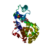 7xe5C 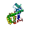 7xe6C 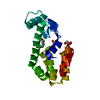 7xe7C  7xe9C  5vnrS C: citing same article ( S: Starting model for refinement |
|---|---|
| Similar structure data |
-Links
 PDBj
PDBj


- Assembly
| Deposited unit | 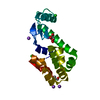
| ||||||||||||
|---|---|---|---|---|---|---|---|---|---|---|---|---|---|
| 1 |
| ||||||||||||
| Unit cell |
|
-Components
| #1: Protein | Mass: 18667.572 Da / Num. of mol.: 1 / Mutation: S44C, C54T, N68C, A93C, C97A, T115C Source method: isolated from a genetically manipulated source Source: (gene. exp.) Escherichia virus T4 / Gene: e, T4Tp126 / Production host:  | ||||||||||
|---|---|---|---|---|---|---|---|---|---|---|---|
| #2: Chemical |   Mass: 92.094 Da / Num. of mol.: 2 / Source method: obtained synthetically / Formula: C3H8O3 Mass: 92.094 Da / Num. of mol.: 2 / Source method: obtained synthetically / Formula: C3H8O3#3: Chemical | ChemComp-NA /   Mass: 22.990 Da / Num. of mol.: 4 / Source method: obtained synthetically / Formula: Na Mass: 22.990 Da / Num. of mol.: 4 / Source method: obtained synthetically / Formula: Na#4: Chemical |   Mass: 35.453 Da / Num. of mol.: 2 / Source method: obtained synthetically / Formula: Cl Mass: 35.453 Da / Num. of mol.: 2 / Source method: obtained synthetically / Formula: Cl#5: Water | ChemComp-HOH / |  Mass: 18.015 Da / Num. of mol.: 270 / Source method: isolated from a natural source / Formula: H2O Mass: 18.015 Da / Num. of mol.: 270 / Source method: isolated from a natural source / Formula: H2OHas ligand of interest | N | Has protein modification | Y | |
-Experimental details
-Experiment
| Experiment | Method: X-RAY DIFFRACTION / Number of used crystals: 1 |
|---|
- Sample preparation
| Crystal | Density Matthews: 2.68 Å3/Da / Density % sol: 54.18 % |
|---|---|
| Crystal grow | Temperature: 293 K / Method: vapor diffusion Details: A crystal was soaked into the precipitant-free solutions containing 40% DMSO, and then backsoaked into the precipitant-free solution. |
-Data collection
| Diffraction | Mean temperature: 100 K / Serial crystal experiment: N |
|---|---|
| Diffraction source | Source: SYNCHROTRON / Site: Photon Factory  / Beamline: BL-5A / Wavelength: 1 Å / Beamline: BL-5A / Wavelength: 1 Å |
| Detector | Type: ADSC QUANTUM 315 / Detector: CCD / Date: Nov 6, 2007 |
| Radiation | Protocol: SINGLE WAVELENGTH / Monochromatic (M) / Laue (L): M / Scattering type: x-ray |
| Radiation wavelength | Wavelength: 1 Å / Relative weight: 1 |
| Reflection | Resolution: 1.1→35.4 Å / Num. obs: 81025 / % possible obs: 98.7 % / Redundancy: 9.8 % / Biso Wilson estimate: 11.94 Å2 / CC1/2: 0.999 / Rrim(I) all: 0.062 / Net I/σ(I): 23.3 |
| Reflection shell | Resolution: 1.1→1.14 Å / Redundancy: 5.5 % / Mean I/σ(I) obs: 2.1 / Num. unique obs: 7157 / CC1/2: 0.812 / Rrim(I) all: 0.63 / % possible all: 88.4 |
- Processing
| Software |
| |||||||||||||||||||||||||||||||||||||||||||||||||||||||||||||||||||||||||||||||||||||||||||||||||||||||||
|---|---|---|---|---|---|---|---|---|---|---|---|---|---|---|---|---|---|---|---|---|---|---|---|---|---|---|---|---|---|---|---|---|---|---|---|---|---|---|---|---|---|---|---|---|---|---|---|---|---|---|---|---|---|---|---|---|---|---|---|---|---|---|---|---|---|---|---|---|---|---|---|---|---|---|---|---|---|---|---|---|---|---|---|---|---|---|---|---|---|---|---|---|---|---|---|---|---|---|---|---|---|---|---|---|---|---|
| Refinement | Method to determine structure: MOLECULAR REPLACEMENT Starting model: 5VNR Resolution: 1.1→25.05 Å / SU ML: 0.0875 / Cross valid method: FREE R-VALUE / σ(F): 1.34 / Phase error: 18.7436 Stereochemistry target values: GeoStd + Monomer Library + CDL v1.2
| |||||||||||||||||||||||||||||||||||||||||||||||||||||||||||||||||||||||||||||||||||||||||||||||||||||||||
| Solvent computation | Shrinkage radii: 0.9 Å / VDW probe radii: 1.1 Å / Solvent model: FLAT BULK SOLVENT MODEL | |||||||||||||||||||||||||||||||||||||||||||||||||||||||||||||||||||||||||||||||||||||||||||||||||||||||||
| Displacement parameters | Biso mean: 18.84 Å2 | |||||||||||||||||||||||||||||||||||||||||||||||||||||||||||||||||||||||||||||||||||||||||||||||||||||||||
| Refinement step | Cycle: LAST / Resolution: 1.1→25.05 Å
| |||||||||||||||||||||||||||||||||||||||||||||||||||||||||||||||||||||||||||||||||||||||||||||||||||||||||
| Refine LS restraints |
| |||||||||||||||||||||||||||||||||||||||||||||||||||||||||||||||||||||||||||||||||||||||||||||||||||||||||
| LS refinement shell |
|
