 Movie
Movie Controller

[English] 日本語
 Yorodumi
- PDB-7xe8: Crystal structure of imine reductase from Streptomyces albidoflavus -
+ Open data

- Basic information
| Entry | Database: PDB / ID: 7xe8 | ||||||
|---|---|---|---|---|---|---|---|
| Title | Crystal structure of imine reductase from Streptomyces albidoflavus | ||||||
 Components Components | 6-phosphogluconate dehydrogenase NAD-binding | ||||||
 Keywords Keywords | OXIDOREDUCTASE / Streptomyces albidoflavus / NADP / R-imine reductase | ||||||
| Function / homology | 3-hydroxyisobutyrate dehydrogenase-related / 6-phosphogluconate dehydrogenase, NADP-binding / NAD binding domain of 6-phosphogluconate dehydrogenase / 6-phosphogluconate dehydrogenase, domain 2 / NADP binding / oxidoreductase activity / NAD(P)-binding domain superfamily / 6-phosphogluconate dehydrogenase NAD-binding Function and homology information Function and homology information | ||||||
| Biological species |  Streptomyces albidoflavus (bacteria) Streptomyces albidoflavus (bacteria) | ||||||
| Method |  X-RAY DIFFRACTION / SYNCHROTRON / MOLECULAR REPLACEMENT / Resolution: 1.72 Å X-RAY DIFFRACTION / SYNCHROTRON / MOLECULAR REPLACEMENT / Resolution: 1.72 Å | ||||||
 Authors Authors | Zhang, J. / Chen, R.C. / Gao, S.S. | ||||||
| Funding support |  China, 1items China, 1items
| ||||||
 Citation Citation | Journal: Commun Chem / Year: 2022 Title: Actinomycetes-derived imine reductases with a preference towards bulky amine substrates Authors: Zhang, J. / Li, X. / Chen, R. / Tan, X. / Liu, X. / Ma, Y. / Zhu, F. / An, C. / Wei, G. / Yao, Y. / Yang, L. / Zhang, P. / Wu, Q. / Sun, Z. / Wang, B.G. / Gao, S.S. / Cui, C. | ||||||
| History |
|
- Structure visualization
| Structure viewer | Molecule: MolmilJmol/JSmol |
|---|
- Downloads & links
-Download
| PDBx/mmCIF format | 7xe8.cif.gz | 225.1 KB | Display | PDBx/mmCIF format |
|---|---|---|---|---|
| PDB format | pdb7xe8.ent.gz | 143.1 KB | Display | PDB format |
| PDBx/mmJSON format | 7xe8.json.gz | Tree view | PDBx/mmJSON format | |
| Others |  Other downloads Other downloads |
-Validation report
| Arichive directory | https://data.pdbj.org/pub/pdb/validation_reports/xe/7xe8ftp://data.pdbj.org/pub/pdb/validation_reports/xe/7xe8 | HTTPS FTP |
|---|
-Related structure data
| Related structure data |  3zgyS S: Starting model for refinement |
|---|---|
| Similar structure data |
-Links
 PDBj
PDBj
- Assembly
| Deposited unit | 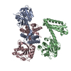
| ||||||||||||
|---|---|---|---|---|---|---|---|---|---|---|---|---|---|
| 1 | 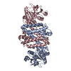
| ||||||||||||
| 2 | 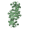
| ||||||||||||
| Unit cell |
|
-Components
| #1: Protein | Mass: 32428.541 Da / Num. of mol.: 3 Source method: isolated from a genetically manipulated source Source: (gene. exp.) Streptomyces albidoflavus (bacteria) / Gene: SSHG_01979, XNRR2_3955 / Production host: #2: Water | ChemComp-HOH / |  Mass: 18.015 Da / Num. of mol.: 884 / Source method: isolated from a natural source / Formula: H2O Mass: 18.015 Da / Num. of mol.: 884 / Source method: isolated from a natural source / Formula: H2O |
|---|
-Experimental details
-Experiment
| Experiment | Method: X-RAY DIFFRACTION / Number of used crystals: 1 |
|---|
- Sample preparation
| Crystal | Density Matthews: 2.48 Å3/Da / Density % sol: 50.4 % |
|---|---|
| Crystal grow | Temperature: 293 K / Method: vapor diffusion, hanging drop / pH: 5.5 Details: 0.2M ammonium acetate, 0.1M Bis Tris pH5.5, 25% PEG 3350 |
-Data collection
| Diffraction | Mean temperature: 100 K / Serial crystal experiment: N |
|---|---|
| Diffraction source | Source: SYNCHROTRON / Site: SSRF / Beamline: BL17U1 / Wavelength: 0.97918 Å |
| Detector | Type: DECTRIS EIGER X 16M / Detector: PIXEL / Date: Oct 24, 2020 |
| Radiation | Protocol: SINGLE WAVELENGTH / Monochromatic (M) / Laue (L): M / Scattering type: x-ray |
| Radiation wavelength | Wavelength: 0.97918 Å / Relative weight: 1 |
| Reflection | Resolution: 1.72→27.07 Å / Num. obs: 90868 / % possible obs: 96.62 % / Redundancy: 6.4 % / Biso Wilson estimate: 25.84 Å2 / CC1/2: 1 / Rmerge(I) obs: 0.03601 / Rpim(I) all: 0.01497 / Rrim(I) all: 0.03908 / Net I/σ(I): 28.13 |
| Reflection shell | Resolution: 1.72→1.781 Å / Rmerge(I) obs: 0.4187 / Mean I/σ(I) obs: 2.75 / Num. unique obs: 8782 / CC1/2: 0.2118 |
- Processing
| Software |
| |||||||||||||||||||||||||||||||||||||||||||||||||||||||||||||||||||||||||||||||||||||||||||||||||||||||||
|---|---|---|---|---|---|---|---|---|---|---|---|---|---|---|---|---|---|---|---|---|---|---|---|---|---|---|---|---|---|---|---|---|---|---|---|---|---|---|---|---|---|---|---|---|---|---|---|---|---|---|---|---|---|---|---|---|---|---|---|---|---|---|---|---|---|---|---|---|---|---|---|---|---|---|---|---|---|---|---|---|---|---|---|---|---|---|---|---|---|---|---|---|---|---|---|---|---|---|---|---|---|---|---|---|---|---|
| Refinement | Method to determine structure: MOLECULAR REPLACEMENT Starting model: 3zgy Resolution: 1.72→27.07 Å / SU ML: 0.1679 / Cross valid method: FREE R-VALUE / σ(F): 2 / Phase error: 21.6516 Stereochemistry target values: GeoStd + Monomer Library + CDL v1.2
| |||||||||||||||||||||||||||||||||||||||||||||||||||||||||||||||||||||||||||||||||||||||||||||||||||||||||
| Solvent computation | Shrinkage radii: 0.9 Å / VDW probe radii: 1.11 Å / Solvent model: FLAT BULK SOLVENT MODEL | |||||||||||||||||||||||||||||||||||||||||||||||||||||||||||||||||||||||||||||||||||||||||||||||||||||||||
| Displacement parameters | Biso mean: 28.91 Å2 | |||||||||||||||||||||||||||||||||||||||||||||||||||||||||||||||||||||||||||||||||||||||||||||||||||||||||
| Refinement step | Cycle: LAST / Resolution: 1.72→27.07 Å
| |||||||||||||||||||||||||||||||||||||||||||||||||||||||||||||||||||||||||||||||||||||||||||||||||||||||||
| Refine LS restraints |
| |||||||||||||||||||||||||||||||||||||||||||||||||||||||||||||||||||||||||||||||||||||||||||||||||||||||||
| LS refinement shell |
|
