National Natural Science Foundation of China (NSFC)
China
Citation
Journal: Nature / Year: 2023 Title: Structural and mechanistic insights into fungal β-1,3-glucan synthase FKS1. Authors: Xinlin Hu / Ping Yang / Changdong Chai / Jia Liu / Huanhuan Sun / Yanan Wu / Mingjie Zhang / Min Zhang / Xiaotian Liu / Hongjun Yu / Abstract: The membrane-integrated synthase FKS is involved in the biosynthesis of β-1,3-glucan, the core component of the fungal cell wall. FKS is the target of widely prescribed antifungal drugs, including ...The membrane-integrated synthase FKS is involved in the biosynthesis of β-1,3-glucan, the core component of the fungal cell wall. FKS is the target of widely prescribed antifungal drugs, including echinocandin and ibrexafungerp. Unfortunately, the mechanism of action of FKS remains enigmatic and this has hampered development of more effective medicines targeting the enzyme. Here we present the cryo-electron microscopy structures of Saccharomyces cerevisiae FKS1 and the echinocandin-resistant mutant FKS1(S643P). These structures reveal the active site of the enzyme at the membrane-cytoplasm interface and a glucan translocation path spanning the membrane bilayer. Multiple bound lipids and notable membrane distortions are observed in the FKS1 structures, suggesting active FKS1-membrane interactions. Echinocandin-resistant mutations are clustered at a region near TM5-6 and TM8 of FKS1. The structure of FKS1(S643P) reveals altered lipid arrangements in this region, suggesting a drug-resistant mechanism of the mutant enzyme. The structures, the catalytic mechanism and the molecular insights into drug-resistant mutations of FKS1 revealed in this study advance the mechanistic understanding of fungal β-1,3-glucan biosynthesis and establish a foundation for developing new antifungal drugs by targeting FKS.
History
Deposition
Mar 29, 2022
Deposition site: PDBJ / Processing site: PDBJ
Revision 1.0
Mar 29, 2023
Provider: repository / Type: Initial release
Revision 1.0
Mar 29, 2023
Data content type: EM metadata / Data content type: EM metadata / Provider: repository / Type: Initial release
Revision 1.0
Mar 29, 2023
Data content type: FSC / Data content type: FSC / Provider: repository / Type: Initial release
Revision 1.0
Mar 29, 2023
Data content type: Image / Data content type: Image / Provider: repository / Type: Initial release
Revision 1.0
Mar 29, 2023
Data content type: Primary map / Data content type: Primary map / Provider: repository / Type: Initial release
Revision 1.0
Mar 29, 2023
Data content type: FSC / Data content type: FSC / Provider: repository / Type: Initial release
Revision 1.0
Mar 29, 2023
Data content type: Image / Data content type: Image / Provider: repository / Type: Initial release
Revision 1.0
Mar 29, 2023
Data content type: Primary map / Data content type: Primary map / Provider: repository / Type: Initial release
Data content type: EM metadata / Data content type: EM metadata / EM metadata / Group: Data processing / Experimental summary / Data content type: EM metadata / EM metadata / Category: em_admin / em_software / Data content type: EM metadata / EM metadata / Item: _em_admin.last_update / _em_software.name
1,3-beta-glucansynthasecomponentFKS1 / 1 / 3-beta-D-glucan-UDP glucosyltransferase / Calcineurin dependent protein 1 / Calcofluor white ...1 / 3-beta-D-glucan-UDP glucosyltransferase / Calcineurin dependent protein 1 / Calcofluor white hypersensitivity protein 53 / Echinocandin target gene protein 1 / FK506 sensitivity protein 1 / Glucan synthase of cerevisiae protein 1 / Papulacandin B resistance protein 1
Mass: 215076.156 Da / Num. of mol.: 1 / Source method: isolated from a natural source / Source: (natural) Saccharomyces cerevisiae (brewer's yeast) / Strain: ATCC 204508 / S288c / References: UniProt: P38631, 1,3-beta-glucan synthase
In the structure databanks used in Yorodumi, some data are registered as the other names, "COVID-19 virus" and "2019-nCoV". Here are the details of the virus and the list of structure data.
Jan 31, 2019. EMDB accession codes are about to change! (news from PDBe EMDB page)
EMDB accession codes are about to change! (news from PDBe EMDB page)
The allocation of 4 digits for EMDB accession codes will soon come to an end. Whilst these codes will remain in use, new EMDB accession codes will include an additional digit and will expand incrementally as the available range of codes is exhausted. The current 4-digit format prefixed with “EMD-” (i.e. EMD-XXXX) will advance to a 5-digit format (i.e. EMD-XXXXX), and so on. It is currently estimated that the 4-digit codes will be depleted around Spring 2019, at which point the 5-digit format will come into force.
The EM Navigator/Yorodumi systems omit the EMD- prefix.
Related info.:Q: What is EMD? / ID/Accession-code notation in Yorodumi/EM Navigator
Yorodumi is a browser for structure data from EMDB, PDB, SASBDB, etc.
This page is also the successor to EM Navigator detail page, and also detail information page/front-end page for Omokage search.
The word "yorodu" (or yorozu) is an old Japanese word meaning "ten thousand". "mi" (miru) is to see.
Related info.:EMDB / PDB / SASBDB / Comparison of 3 databanks / Yorodumi Search / Aug 31, 2016. New EM Navigator & Yorodumi / Yorodumi Papers / Jmol/JSmol / Function and homology information / Changes in new EM Navigator and Yorodumi
 Movie
Movie Controller


 Open data
 Basic information
 Components
 Keywords
 Function and homology information
 Authors
 China, 1items
  Citation
 Structure visualization
 Downloads & links
 Other downloads

 PDBj
PDBj Assembly
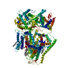


 Mass: 128.255 Da / Num. of mol.: 7 / Source method: obtained synthetically / Formula: C9H20
 Mass: 142.282 Da / Num. of mol.: 6 / Source method: obtained synthetically / Formula: C10H22
 Mass: 198.388 Da / Num. of mol.: 1 / Source method: obtained synthetically / Formula: C14H30
 Mass: 100.202 Da / Num. of mol.: 7 / Source method: obtained synthetically / Formula: C7H16
 Mass: 170.335 Da / Num. of mol.: 3 / Source method: obtained synthetically / Formula: C12H26
 Mass: 495.587 Da / Num. of mol.: 1 / Source method: obtained synthetically / Formula: C23H46NO8P
 Sample preparation
 Electron microscopy imaging
 FIELD EMISSION GUN / Accelerating voltage: 300 kV / Illumination mode: FLOOD BEAM
 Processing