 Movie
Movie Controller

[English] 日本語
 Yorodumi
- PDB-7xb5: Structure of the ligand-binding domain of S. cerevisiae Upc2 in f... -
+ Open data

- Basic information
| Entry | Database: PDB / ID: 7xb5 | ||||||
|---|---|---|---|---|---|---|---|
| Title | Structure of the ligand-binding domain of S. cerevisiae Upc2 in fusion with T4 lysozyme | ||||||
 Components Components | fusion protein of Sterol uptake control protein 2 and Endolysin | ||||||
 Keywords Keywords | TRANSCRIPTION / ergosterol / transcription factor / ligand-binding / zinc finger | ||||||
| Function / homology |  Function and homology information Function and homology informationpositive regulation of sterol import / positive regulation of ergosterol biosynthetic process / positive regulation of filamentous growth of a population of unicellular organisms in response to starvation / viral release from host cell by cytolysis / peptidoglycan catabolic process / cell wall macromolecule catabolic process / lysozyme / lysozyme activity / DNA-binding transcription activator activity, RNA polymerase II-specific / cellular response to hypoxia ...positive regulation of sterol import / positive regulation of ergosterol biosynthetic process / positive regulation of filamentous growth of a population of unicellular organisms in response to starvation / viral release from host cell by cytolysis / peptidoglycan catabolic process / cell wall macromolecule catabolic process / lysozyme / lysozyme activity / DNA-binding transcription activator activity, RNA polymerase II-specific / cellular response to hypoxia / host cell cytoplasm / defense response to bacterium / RNA polymerase II cis-regulatory region sequence-specific DNA binding / regulation of transcription by RNA polymerase II / perinuclear region of cytoplasm / positive regulation of transcription by RNA polymerase II / zinc ion binding / membrane / nucleus / cytoplasm Similarity search - Function | ||||||
| Biological species |   Escherichia virus T4 Escherichia virus T4 | ||||||
| Method |  X-RAY DIFFRACTION / SYNCHROTRON / MOLECULAR REPLACEMENT / Resolution: 3.44 Å X-RAY DIFFRACTION / SYNCHROTRON / MOLECULAR REPLACEMENT / Resolution: 3.44 Å | ||||||
 Authors Authors | Tan, L. / Im, Y.J. | ||||||
| Funding support |  Korea, Republic Of, 1items Korea, Republic Of, 1items
| ||||||
 Citation Citation | Journal: Nat.Chem.Biol. / Year: 2022 Title: Structural basis for activation of fungal sterol receptor Upc2 and azole resistance. Authors: Tan, L. / Chen, L. / Yang, H. / Jin, B. / Kim, G. / Im, Y.J. | ||||||
| History |
|
- Structure visualization
| Structure viewer | Molecule: MolmilJmol/JSmol |
|---|
- Downloads & links
-Download
| PDBx/mmCIF format | 7xb5.cif.gz | 192.2 KB | Display | PDBx/mmCIF format |
|---|---|---|---|---|
| PDB format | pdb7xb5.ent.gz | 128.6 KB | Display | PDB format |
| PDBx/mmJSON format | 7xb5.json.gz | Tree view | PDBx/mmJSON format | |
| Others |  Other downloads Other downloads |
-Validation report
| Arichive directory | https://data.pdbj.org/pub/pdb/validation_reports/xb/7xb5ftp://data.pdbj.org/pub/pdb/validation_reports/xb/7xb5 | HTTPS FTP |
|---|
-Related structure data
| Related structure data | 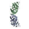 7vprC  7vpsC  7vptC 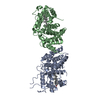 7vpuC  4n9nS S: Starting model for refinement C: citing same article ( |
|---|---|
| Similar structure data |
-Links
 PDBj
PDBj


- Assembly
| Deposited unit | 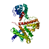
| ||||||||||||
|---|---|---|---|---|---|---|---|---|---|---|---|---|---|
| 1 | 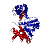
| ||||||||||||
| Unit cell |
|
-Components
| #1: Protein | Mass: 52253.812 Da / Num. of mol.: 1 / Mutation: R727G,C769T,C812A,I852R,I925G Source method: isolated from a genetically manipulated source Details: The loop residues 715-725 of Upc2 were replaced by bacteriophage T4 Lysozyme (residues 2 - 161) using Val-Asp dipeptide as linkers. Source: (gene. exp.) Escherichia virus T4Strain: S288c / Gene: UPC2, MOX4, YDR213W, YD8142.14, YD8142B.05, UPC2 / Plasmid: pET28b / Production host:  |
|---|
-Experimental details
-Experiment
| Experiment | Method: X-RAY DIFFRACTION / Number of used crystals: 1 |
|---|
- Sample preparation
| Crystal | Density Matthews: 3.17 Å3/Da / Density % sol: 61.22 % |
|---|---|
| Crystal grow | Temperature: 296 K / Method: vapor diffusion, hanging drop / pH: 7 Details: 0.1 M HEPES pH 7.0, 12.5% PEG 8000, 0.2 M sodium citrate |
-Data collection
| Diffraction | Mean temperature: 110 K / Serial crystal experiment: N |
|---|---|
| Diffraction source | Source: SYNCHROTRON / Site: PAL/PLS / Beamline: 7A (6B, 6C1) / Wavelength: 0.97934 Å |
| Detector | Type: ADSC QUANTUM 270 / Detector: CCD / Date: Aug 4, 2015 |
| Radiation | Protocol: SINGLE WAVELENGTH / Monochromatic (M) / Laue (L): M / Scattering type: x-ray |
| Radiation wavelength | Wavelength: 0.97934 Å / Relative weight: 1 |
| Reflection | Resolution: 3.44→50 Å / Num. obs: 8514 / % possible obs: 97.9 % / Redundancy: 7.2 % / Biso Wilson estimate: 105.93 Å2 / Rmerge(I) obs: 0.098 / Net I/σ(I): 32.8 |
| Reflection shell | Resolution: 3.5→3.56 Å / Redundancy: 7.1 % / Rmerge(I) obs: 0.561 / Mean I/σ(I) obs: 4.8 / Num. unique obs: 418 / % possible all: 96.5 |
- Processing
| Software |
| ||||||||||||||||||||||||||||||||||||||||
|---|---|---|---|---|---|---|---|---|---|---|---|---|---|---|---|---|---|---|---|---|---|---|---|---|---|---|---|---|---|---|---|---|---|---|---|---|---|---|---|---|---|
| Refinement | Method to determine structure: MOLECULAR REPLACEMENT Starting model: 4N9N Resolution: 3.44→32.98 Å / SU ML: 0.5063 / Cross valid method: FREE R-VALUE / σ(F): 1.34 / Phase error: 34.0065 Stereochemistry target values: GeoStd + Monomer Library + CDL v1.2
| ||||||||||||||||||||||||||||||||||||||||
| Solvent computation | Shrinkage radii: 0.9 Å / VDW probe radii: 1.11 Å / Solvent model: FLAT BULK SOLVENT MODEL | ||||||||||||||||||||||||||||||||||||||||
| Displacement parameters | Biso mean: 120.53 Å2 | ||||||||||||||||||||||||||||||||||||||||
| Refinement step | Cycle: LAST / Resolution: 3.44→32.98 Å
| ||||||||||||||||||||||||||||||||||||||||
| Refine LS restraints |
| ||||||||||||||||||||||||||||||||||||||||
| LS refinement shell |
| ||||||||||||||||||||||||||||||||||||||||
| Refinement TLS params. | Method: refined / Origin x: -4.47222941247 Å / Origin y: -20.5726666228 Å / Origin z: 20.2193100025 Å
| ||||||||||||||||||||||||||||||||||||||||
| Refinement TLS group | Selection details: all |
