 Movie
Movie Controller


+ Open data

- Basic information
| Entry | Database: PDB / ID: 7x8h | ||||||
|---|---|---|---|---|---|---|---|
| Title | Crystal structure of AtHPPD-(+)-Usnic acid complex | ||||||
 Components Components | 4-hydroxyphenylpyruvate dioxygenase | ||||||
 Keywords Keywords | OXIDOREDUCTASE / complex / substrate | ||||||
| Function / homology |  Function and homology information Function and homology informationvitamin E biosynthetic process / 4-hydroxyphenylpyruvate dioxygenase / plastoquinone biosynthetic process / 4-hydroxyphenylpyruvate dioxygenase activity / carotenoid biosynthetic process / L-tyrosine catabolic process / L-phenylalanine catabolic process / chloroplast / iron ion binding / mitochondrion ...vitamin E biosynthetic process / 4-hydroxyphenylpyruvate dioxygenase / plastoquinone biosynthetic process / 4-hydroxyphenylpyruvate dioxygenase activity / carotenoid biosynthetic process / L-tyrosine catabolic process / L-phenylalanine catabolic process / chloroplast / iron ion binding / mitochondrion / extracellular region / identical protein binding / cytosol Similarity search - Function | ||||||
| Biological species |  | ||||||
| Method |  X-RAY DIFFRACTION / SYNCHROTRON / MOLECULAR REPLACEMENT / Resolution: 1.986 Å X-RAY DIFFRACTION / SYNCHROTRON / MOLECULAR REPLACEMENT / Resolution: 1.986 Å | ||||||
 Authors Authors | Dong, J. / Lin, H.-Y. / Yang, G.-F. | ||||||
| Funding support |  China, 1items China, 1items
| ||||||
 Citation Citation | Journal: J.Agric.Food Chem. / Year: 2023 Title: Discovery of Subnanomolar Inhibitors of 4-Hydroxyphenylpyruvate Dioxygenase via Structure-Based Rational Design. Authors: Dong, J. / Dong, J. / Yu, X.H. / Yan, Y.C. / Nan, J.X. / Ye, B.Q. / Yang, W.C. / Lin, H.Y. / Yang, G.F. | ||||||
| History |
|
- Structure visualization
| Structure viewer | Molecule: MolmilJmol/JSmol |
|---|
- Downloads & links
-Download
| PDBx/mmCIF format | 7x8h.cif.gz | 93.6 KB | Display | PDBx/mmCIF format |
|---|---|---|---|---|
| PDB format | pdb7x8h.ent.gz | 66.5 KB | Display | PDB format |
| PDBx/mmJSON format | 7x8h.json.gz | Tree view | PDBx/mmJSON format | |
| Others |  Other downloads Other downloads |
-Validation report
| Arichive directory | https://data.pdbj.org/pub/pdb/validation_reports/x8/7x8hftp://data.pdbj.org/pub/pdb/validation_reports/x8/7x8h | HTTPS FTP |
|---|
-Related structure data
| Related structure data |  7x5yC  7x5zC  7x62C  7x64C  7x67C  7x69C  7x8iC  7xvhC  8howC  7cqsS S: Starting model for refinement C: citing same article ( |
|---|---|
| Similar structure data |
-Links
 PDBj
PDBj
- Assembly
| Deposited unit | 
| ||||||||
|---|---|---|---|---|---|---|---|---|---|
| 1 | 
| ||||||||
| Unit cell |
|
-Components
| #1: Protein | Mass: 45952.727 Da / Num. of mol.: 1 Source method: isolated from a genetically manipulated source Source: (gene. exp.) Production host:  References: UniProt: P93836, 4-hydroxyphenylpyruvate dioxygenase |
|---|---|
| #2: Chemical | ChemComp-CO /   Mass: 58.933 Da / Num. of mol.: 1 / Source method: obtained synthetically / Formula: Co Mass: 58.933 Da / Num. of mol.: 1 / Source method: obtained synthetically / Formula: Co |
| #3: Chemical | ChemComp-AIY / (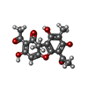  Mass: 344.315 Da / Num. of mol.: 1 / Source method: isolated from a natural source / Formula: C18H16O7 Mass: 344.315 Da / Num. of mol.: 1 / Source method: isolated from a natural source / Formula: C18H16O7 |
| #4: Water | ChemComp-HOH /  Mass: 18.015 Da / Num. of mol.: 127 / Source method: isolated from a natural source / Formula: H2O Mass: 18.015 Da / Num. of mol.: 127 / Source method: isolated from a natural source / Formula: H2O |
| Has ligand of interest | N |
| Has protein modification | Y |
-Experimental details
-Experiment
| Experiment | Method: X-RAY DIFFRACTION / Number of used crystals: 1 |
|---|
- Sample preparation
| Crystal | Density Matthews: 2.27 Å3/Da / Density % sol: 45.78 % |
|---|---|
| Crystal grow | Temperature: 293 K / Method: vapor diffusion, hanging drop Details: 0.1M Tris/Bicine pH 8.5, 15% (v/v) MPD, 15% (w/v) PEG 1000, 15% (w/v) PEG 3350, 0.03M NaBr, 0.03M NaF, 0.03M NaI |
-Data collection
| Diffraction | Mean temperature: 100 K / Serial crystal experiment: N |
|---|---|
| Diffraction source | Source: SYNCHROTRON / Site: SSRF / Beamline: BL19U1 / Wavelength: 1.06 Å |
| Detector | Type: ADSC QUANTUM 315r / Detector: CCD / Date: Aug 8, 2020 |
| Radiation | Protocol: SINGLE WAVELENGTH / Monochromatic (M) / Laue (L): M / Scattering type: x-ray |
| Radiation wavelength | Wavelength: 1.06 Å / Relative weight: 1 |
| Reflection | Resolution: 1.986→30 Å / Num. obs: 25936 / % possible obs: 99.1 % / Redundancy: 6.1 % / CC1/2: 0.99 / Rmerge(I) obs: 0.081 / Net I/σ(I): 21 |
| Reflection shell | Resolution: 2→2.03 Å / Rmerge(I) obs: 0.462 / Num. unique obs: 1268 / CC1/2: 0.914 |
- Processing
| Software |
| ||||||||||||||||||||||||||||||||||||||||||||||||||||||||||||||||||||||
|---|---|---|---|---|---|---|---|---|---|---|---|---|---|---|---|---|---|---|---|---|---|---|---|---|---|---|---|---|---|---|---|---|---|---|---|---|---|---|---|---|---|---|---|---|---|---|---|---|---|---|---|---|---|---|---|---|---|---|---|---|---|---|---|---|---|---|---|---|---|---|---|
| Refinement | Method to determine structure: MOLECULAR REPLACEMENT Starting model: 7CQS Resolution: 1.986→29.206 Å / SU ML: 0.19 / Cross valid method: FREE R-VALUE / σ(F): 1.38 / Phase error: 21.04 / Stereochemistry target values: ML
| ||||||||||||||||||||||||||||||||||||||||||||||||||||||||||||||||||||||
| Solvent computation | Shrinkage radii: 0.9 Å / VDW probe radii: 1.11 Å / Solvent model: FLAT BULK SOLVENT MODEL | ||||||||||||||||||||||||||||||||||||||||||||||||||||||||||||||||||||||
| Refinement step | Cycle: LAST / Resolution: 1.986→29.206 Å
| ||||||||||||||||||||||||||||||||||||||||||||||||||||||||||||||||||||||
| Refine LS restraints |
| ||||||||||||||||||||||||||||||||||||||||||||||||||||||||||||||||||||||
| LS refinement shell |
|
