 Movie
Movie Controller

[English] 日本語
 Yorodumi
- PDB-7x7w: The X-ray Crystallographic Structure of D-Psicose 3-epimerase fro... -
+ Open data

- Basic information
| Entry | Database: PDB / ID: 7x7w | ||||||||||||
|---|---|---|---|---|---|---|---|---|---|---|---|---|---|
| Title | The X-ray Crystallographic Structure of D-Psicose 3-epimerase from Clostridia bacterium | ||||||||||||
 Components Components | D-PSICOSE 3-EPIMERASE | ||||||||||||
 Keywords Keywords | ISOMERASE / D-Psicose 3-epimerase | ||||||||||||
| Function / homology | :  Function and homology information Function and homology information | ||||||||||||
| Biological species |  Clostridia bacterium (bacteria) Clostridia bacterium (bacteria) | ||||||||||||
| Method |  X-RAY DIFFRACTION / SYNCHROTRON / MOLECULAR REPLACEMENT / Resolution: 2.097 Å X-RAY DIFFRACTION / SYNCHROTRON / MOLECULAR REPLACEMENT / Resolution: 2.097 Å | ||||||||||||
 Authors Authors | Li, Z.F. / Ban, X.F. / Xie, X.F. / Tian, Y.X. / Li, C.M. / Gu, Z.B. | ||||||||||||
| Funding support |  China, 3items China, 3items
| ||||||||||||
 Citation Citation | Journal: Acta Crystallogr D Struct Biol / Year: 2022 Title: Crystal structure of a novel homodimeric D-allulose 3-epimerase from a Clostridia bacterium. Authors: Xie, X. / Tian, Y. / Ban, X. / Li, C. / Yang, H. / Li, Z. | ||||||||||||
| History |
|
- Structure visualization
| Structure viewer | Molecule: MolmilJmol/JSmol |
|---|
- Downloads & links
-Download
| PDBx/mmCIF format | 7x7w.cif.gz | 125.9 KB | Display | PDBx/mmCIF format |
|---|---|---|---|---|
| PDB format | pdb7x7w.ent.gz | 97.8 KB | Display | PDB format |
| PDBx/mmJSON format | 7x7w.json.gz | Tree view | PDBx/mmJSON format | |
| Others |  Other downloads Other downloads |
-Validation report
| Arichive directory | https://data.pdbj.org/pub/pdb/validation_reports/x7/7x7wftp://data.pdbj.org/pub/pdb/validation_reports/x7/7x7w | HTTPS FTP |
|---|
-Related structure data
| Related structure data |  3vnkS S: Starting model for refinement |
|---|---|
| Similar structure data |
-Links
 PDBj
PDBj- Assembly
| Deposited unit | 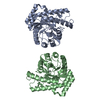
| ||||||||||
|---|---|---|---|---|---|---|---|---|---|---|---|
| 1 | 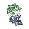
| ||||||||||
| Unit cell |
| ||||||||||
| Components on special symmetry positions |
|
-Components
| #1: Protein | Mass: 32348.537 Da / Num. of mol.: 2 Source method: isolated from a genetically manipulated source Source: (gene. exp.) Clostridia bacterium (bacteria) / Production host: #2: Chemical |   Mass: 54.938 Da / Num. of mol.: 2 / Source method: isolated from a natural source / Formula: Mn / Feature type: SUBJECT OF INVESTIGATION Mass: 54.938 Da / Num. of mol.: 2 / Source method: isolated from a natural source / Formula: Mn / Feature type: SUBJECT OF INVESTIGATION#3: Water | ChemComp-HOH / |  Mass: 18.015 Da / Num. of mol.: 161 / Source method: isolated from a natural source / Formula: H2O Mass: 18.015 Da / Num. of mol.: 161 / Source method: isolated from a natural source / Formula: H2OHas ligand of interest | Y | Has protein modification | N | |
|---|
-Experimental details
-Experiment
| Experiment | Method: X-RAY DIFFRACTION / Number of used crystals: 1 |
|---|
- Sample preparation
| Crystal | Density Matthews: 2.46 Å3/Da / Density % sol: 49.96 % |
|---|---|
| Crystal grow | Temperature: 293 K / Method: vapor diffusion, hanging drop Details: 0.1 M Succine acid pH 7.0, 0.1 M HEPES pH7.0, 1% w/v PEG MME 2000 |
-Data collection
| Diffraction | Mean temperature: 100 K / Serial crystal experiment: N |
|---|---|
| Diffraction source | Source: SYNCHROTRON / Site: SSRF / Beamline: BL19U1 / Wavelength: 0.97915 Å |
| Detector | Type: DECTRIS PILATUS 6M / Detector: PIXEL / Date: Dec 9, 2021 |
| Radiation | Protocol: SINGLE WAVELENGTH / Monochromatic (M) / Laue (L): M / Scattering type: x-ray |
| Radiation wavelength | Wavelength: 0.97915 Å / Relative weight: 1 |
| Reflection | Resolution: 2.09→37.95 Å / Num. obs: 37135 / % possible obs: 99.4 % / Redundancy: 6.6 % / Biso Wilson estimate: 34.68 Å2 / Rmerge(I) obs: 0.032 / Net I/σ(I): 30.4 |
| Reflection shell | Resolution: 2.1→2.16 Å / Rmerge(I) obs: 0.124 / Num. unique obs: 2994 |
- Processing
| Software |
| ||||||||||||||||||||||||||||||||||||||||||||||||||||||||||||||||||||||||||||||||||||||||||
|---|---|---|---|---|---|---|---|---|---|---|---|---|---|---|---|---|---|---|---|---|---|---|---|---|---|---|---|---|---|---|---|---|---|---|---|---|---|---|---|---|---|---|---|---|---|---|---|---|---|---|---|---|---|---|---|---|---|---|---|---|---|---|---|---|---|---|---|---|---|---|---|---|---|---|---|---|---|---|---|---|---|---|---|---|---|---|---|---|---|---|---|
| Refinement | Method to determine structure: MOLECULAR REPLACEMENT Starting model: 3VNK Resolution: 2.097→37.939 Å / SU ML: 0.21 / Cross valid method: THROUGHOUT / σ(F): 1.38 / Phase error: 34.92 / Stereochemistry target values: ML
| ||||||||||||||||||||||||||||||||||||||||||||||||||||||||||||||||||||||||||||||||||||||||||
| Solvent computation | Shrinkage radii: 0.9 Å / VDW probe radii: 1.11 Å / Solvent model: FLAT BULK SOLVENT MODEL | ||||||||||||||||||||||||||||||||||||||||||||||||||||||||||||||||||||||||||||||||||||||||||
| Displacement parameters | Biso max: 96 Å2 / Biso mean: 50.2439 Å2 / Biso min: 27.64 Å2 | ||||||||||||||||||||||||||||||||||||||||||||||||||||||||||||||||||||||||||||||||||||||||||
| Refinement step | Cycle: final / Resolution: 2.097→37.939 Å
| ||||||||||||||||||||||||||||||||||||||||||||||||||||||||||||||||||||||||||||||||||||||||||
| LS refinement shell | Refine-ID: X-RAY DIFFRACTION / Rfactor Rfree error: 0
|
