 Movie
Movie Controller


+ Open data

- Basic information
| Entry | Database: PDB / ID: 7x7m | ||||||||||||||||||||||||||||||||||||||||||||||||||||||
|---|---|---|---|---|---|---|---|---|---|---|---|---|---|---|---|---|---|---|---|---|---|---|---|---|---|---|---|---|---|---|---|---|---|---|---|---|---|---|---|---|---|---|---|---|---|---|---|---|---|---|---|---|---|---|---|
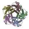
| Title | Lumazine Synthase from Aquifex aeolicus | ||||||||||||||||||||||||||||||||||||||||||||||||||||||
 Components Components | 6,7-dimethyl-8-ribityllumazine synthase | ||||||||||||||||||||||||||||||||||||||||||||||||||||||
 Keywords Keywords | TRANSFERASE / Thermostable Transferase | ||||||||||||||||||||||||||||||||||||||||||||||||||||||
| Function / homology |  Function and homology information Function and homology information6,7-dimethyl-8-ribityllumazine synthase / 6,7-dimethyl-8-ribityllumazine synthase activity / riboflavin synthase complex / riboflavin biosynthetic process / cytoplasm / cytosol Similarity search - Function | ||||||||||||||||||||||||||||||||||||||||||||||||||||||
| Biological species |   Aquifex aeolicus VF5 (bacteria) Aquifex aeolicus VF5 (bacteria) | ||||||||||||||||||||||||||||||||||||||||||||||||||||||
| Method | ELECTRON MICROSCOPY / single particle reconstruction / cryo EM / Resolution: 2.33 Å | ||||||||||||||||||||||||||||||||||||||||||||||||||||||
 Authors Authors | Sobhy, M.A. / Hamdan, S.M. | ||||||||||||||||||||||||||||||||||||||||||||||||||||||
| Funding support |  Saudi Arabia, 1items Saudi Arabia, 1items
| ||||||||||||||||||||||||||||||||||||||||||||||||||||||
 Citation Citation | Journal: PLoS One / Year: 2022 Title: Cryo-electron structures of the extreme thermostable enzymes Sulfur Oxygenase Reductase and Lumazine Synthase. Authors: Mohamed A Sobhy / Lingyun Zhao / Dalaver Anjum / Ali Behzad / Masateru Takahashi / Muhammad Tehseen / Alfredo De Biasio / Rachid Sougrat / Samir Hamdan / Abstract: Thermostable enzymes have the potential for use in a wide variety of biotechnological applications. Cryo-electron microscopy (cryo-EM) enables the imaging of biomolecules in their native aqueous ...Thermostable enzymes have the potential for use in a wide variety of biotechnological applications. Cryo-electron microscopy (cryo-EM) enables the imaging of biomolecules in their native aqueous environment. Here, we present high resolution cryo-EM structures of two thermostable enzymes that exhibit multimeric cage-like structures arranged into two different point-group symmetries. First, we determined the structure of the Sulfur Oxygenase Reductase (SOR) enzyme that catalyzes both the oxygenation and disproportionation of elemental sulfur in Archea and is composed of 24 homomeric units each of MW ≃ 35 kDa arranged in octahedral symmetry. The structure of SOR from Acidianus ambivalens (7X9W) was determined at 2.78 Å resolution. The active site of each subunit inside the central nanocompartment is composed of Fe3+ coordinated to two water molecules and the three amino acids (H86, H90 and E114). Second, we determined the structure of Lumazine Synthase (LS) from Aquifex aeolicus (7X7M) at 2.33 Å resolution. LS forms a cage-like structure consisting of 60 identical subunits each of MW ≃ 15 kDa arranged in a strict icosahedral symmetry. The LS subunits are interconnected by ion-pair network. Due to their thermostability and relatively easy purification scheme, both SOR and LS can serve as a model for the catalytic and structural characterization of biocatalysts as well as a benchmark for cryo-EM sample preparation, optimization of the acquisition parameters and 3D reconstruction. | ||||||||||||||||||||||||||||||||||||||||||||||||||||||
| History |
|
- Structure visualization
| Structure viewer | Molecule: MolmilJmol/JSmol |
|---|
- Downloads & links
-Download
| PDBx/mmCIF format | 7x7m.cif.gz | 1.4 MB | Display | PDBx/mmCIF format |
|---|---|---|---|---|
| PDB format | pdb7x7m.ent.gz | 1.2 MB | Display | PDB format |
| PDBx/mmJSON format | 7x7m.json.gz | Tree view | PDBx/mmJSON format | |
| Others |  Other downloads Other downloads |
-Validation report
| Arichive directory | https://data.pdbj.org/pub/pdb/validation_reports/x7/7x7mftp://data.pdbj.org/pub/pdb/validation_reports/x7/7x7m | HTTPS FTP |
|---|
-Related structure data
| Related structure data | 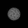 33041MC 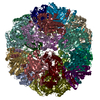 7x9wC M: map data used to model this data C: citing same article ( |
|---|---|
| Similar structure data |

-Links
 PDBj
PDBj- Assembly
| Deposited unit | 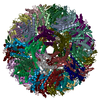
|
|---|---|
| 1 |
|
-Components
| #1: Protein | Mass: 16727.201 Da / Num. of mol.: 60 Source method: isolated from a genetically manipulated source Source: (gene. exp.) Aquifex aeolicus VF5 (bacteria) / Strain: VF5 / Gene: ribH / Production host: References: UniProt: O66529, 6,7-dimethyl-8-ribityllumazine synthase Has protein modification | N | |
|---|
-Experimental details
-Experiment
| Experiment | Method: ELECTRON MICROSCOPY |
|---|---|
| EM experiment | Aggregation state: PARTICLE / 3D reconstruction method: single particle reconstruction |
- Sample preparation
| Component | Name: CryoEM structure of Lumazine synthase composed of 60 identical subunits Type: COMPLEX / Entity ID: all / Source: RECOMBINANT | |||||||||||||||
|---|---|---|---|---|---|---|---|---|---|---|---|---|---|---|---|---|
| Source (natural) | Organism: Aquifex aeolicus (bacteria) | |||||||||||||||
| Source (recombinant) | Organism: | |||||||||||||||
| Buffer solution | pH: 7.5 | |||||||||||||||
| Buffer component |
| |||||||||||||||
| Specimen | Conc.: 6 mg/ml / Embedding applied: NO / Shadowing applied: NO / Staining applied: NO / Vitrification applied: YES | |||||||||||||||
| Specimen support | Grid material: GOLD / Grid mesh size: 300 divisions/in. / Grid type: Quantifoil R2/2 | |||||||||||||||
| Vitrification | Instrument: FEI VITROBOT MARK I / Cryogen name: ETHANE / Humidity: 100 % / Chamber temperature: 295 K Details: The purified protein was applied to the glow discharged grids inside the chamber, blotted for a duration of 1.5 s, and then plunge-frozen in liquid ethane cooled by liquid nitrogen. |
- Electron microscopy imaging
| Experimental equipment |  Model: Titan Krios / Image courtesy: FEI Company |
|---|---|
| Microscopy | Model: FEI TITAN KRIOS |
| Electron gun | Electron source:  FIELD EMISSION GUN / Accelerating voltage: 300 kV / Illumination mode: SPOT SCAN FIELD EMISSION GUN / Accelerating voltage: 300 kV / Illumination mode: SPOT SCAN |
| Electron lens | Mode: BRIGHT FIELD / Nominal magnification: 130000 X / Nominal defocus max: 1200 nm / Nominal defocus min: 200 nm |
| Specimen holder | Specimen holder model: FEI TITAN KRIOS AUTOGRID HOLDER |
| Image recording | Electron dose: 70 e/Å2 / Detector mode: SUPER-RESOLUTION / Film or detector model: GATAN K2 SUMMIT (4k x 4k) |
| Image scans | Movie frames/image: 50 |
- Processing
| Software | Name: PHENIX / Version: 1.18.2_3874: / Classification: refinement | ||||||||||||||||||||||||
|---|---|---|---|---|---|---|---|---|---|---|---|---|---|---|---|---|---|---|---|---|---|---|---|---|---|
| EM software |
| ||||||||||||||||||||||||
| CTF correction | Type: NONE | ||||||||||||||||||||||||
| Particle selection | Num. of particles selected: 344110 Details: particles were selected by LoG picking from 1516 micrographs | ||||||||||||||||||||||||
| Symmetry | Point symmetry: I (icosahedral) | ||||||||||||||||||||||||
| 3D reconstruction | Resolution: 2.33 Å / Resolution method: FSC 0.143 CUT-OFF / Num. of particles: 103328 / Num. of class averages: 1 / Symmetry type: POINT | ||||||||||||||||||||||||
| Refine LS restraints |
|
