 Movie
Movie Controller

[English] 日本語
 Yorodumi
- PDB-7x1x: Crystal Structure of cis-4,5-dihydrodiol phthalate dehydrogenase ... -
+ Open data

- Basic information
| Entry | Database: PDB / ID: 7x1x | ||||||
|---|---|---|---|---|---|---|---|
| Title | Crystal Structure of cis-4,5-dihydrodiol phthalate dehydrogenase in complex with NAD+ | ||||||
 Components Components | 4,5-dihydroxyphthalate dehydrogenase | ||||||
 Keywords Keywords | OXIDOREDUCTASE / dehydrogenase / bacterial protein / NAD cofactor | ||||||
| Function / homology | NICOTINAMIDE-ADENINE-DINUCLEOTIDE Function and homology information Function and homology information | ||||||
| Biological species |  Comamonas testosteroni KF-1 (bacteria) Comamonas testosteroni KF-1 (bacteria) | ||||||
| Method |  X-RAY DIFFRACTION / SYNCHROTRON / MOLECULAR REPLACEMENT / Resolution: 2.77 Å X-RAY DIFFRACTION / SYNCHROTRON / MOLECULAR REPLACEMENT / Resolution: 2.77 Å | ||||||
 Authors Authors | Sharma, M. / Mahto, J.K. / Kumar, P. | ||||||
| Funding support |  India, 1items India, 1items
| ||||||
 Citation Citation | Journal: Arch.Biochem.Biophys. / Year: 2022 Title: Conformational flexibility enables catalysis of phthalate cis-4,5-dihydrodiol dehydrogenase. Authors: Mahto, J.K. / Sharma, M. / Neetu, N. / Kayastha, A. / Aggarwal, S. / Kumar, P. | ||||||
| History |
|
- Structure visualization
| Structure viewer | Molecule: MolmilJmol/JSmol |
|---|
- Downloads & links
-Download
| PDBx/mmCIF format | 7x1x.cif.gz | 286 KB | Display | PDBx/mmCIF format |
|---|---|---|---|---|
| PDB format | pdb7x1x.ent.gz | 230.3 KB | Display | PDB format |
| PDBx/mmJSON format | 7x1x.json.gz | Tree view | PDBx/mmJSON format | |
| Others |  Other downloads Other downloads |
-Validation report
| Arichive directory | https://data.pdbj.org/pub/pdb/validation_reports/x1/7x1xftp://data.pdbj.org/pub/pdb/validation_reports/x1/7x1x | HTTPS FTP |
|---|
-Related structure data
| Related structure data | 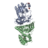 7wzdC  7x2yC  3moiS S: Starting model for refinement C: citing same article ( |
|---|---|
| Similar structure data |
-Links
 PDBj
PDBj- Assembly
| Deposited unit | 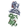
| ||||||||||||||||||
|---|---|---|---|---|---|---|---|---|---|---|---|---|---|---|---|---|---|---|---|
| 1 |
| ||||||||||||||||||
| Unit cell |
| ||||||||||||||||||
| Noncrystallographic symmetry (NCS) | NCS domain:
NCS domain segments: Component-ID: _ / Ens-ID: 1 / Beg auth comp-ID: GLY / Beg label comp-ID: GLY / End auth comp-ID: PRO / End label comp-ID: PRO / Refine code: _ / Auth seq-ID: 4 - 391 / Label seq-ID: 4 - 391
|
-Components
| #1: Protein | Mass: 43046.676 Da / Num. of mol.: 2 Source method: isolated from a genetically manipulated source Source: (gene. exp.) Comamonas testosteroni KF-1 (bacteria) / Gene: O987_21845 / Production host: #2: Chemical |   Mass: 92.094 Da / Num. of mol.: 3 / Source method: obtained synthetically / Formula: C3H8O3 Mass: 92.094 Da / Num. of mol.: 3 / Source method: obtained synthetically / Formula: C3H8O3#3: Chemical |   Mass: 663.425 Da / Num. of mol.: 2 / Source method: obtained synthetically / Formula: C21H27N7O14P2 / Feature type: SUBJECT OF INVESTIGATION / Comment: NAD*YM Mass: 663.425 Da / Num. of mol.: 2 / Source method: obtained synthetically / Formula: C21H27N7O14P2 / Feature type: SUBJECT OF INVESTIGATION / Comment: NAD*YM#4: Water | ChemComp-HOH / |  Mass: 18.015 Da / Num. of mol.: 149 / Source method: isolated from a natural source / Formula: H2O Mass: 18.015 Da / Num. of mol.: 149 / Source method: isolated from a natural source / Formula: H2OHas ligand of interest | Y | |
|---|
-Experimental details
-Experiment
| Experiment | Method: X-RAY DIFFRACTION / Number of used crystals: 1 |
|---|
- Sample preparation
| Crystal | Density Matthews: 2.36 Å3/Da / Density % sol: 47.87 % |
|---|---|
| Crystal grow | Temperature: 293 K / Method: vapor diffusion, sitting drop Details: 20% PEG3350, 0.2M AMMONIUM PHOSPHATE DIBASIC, 100mM TRIS BUFFER pH 8.0 |
-Data collection
| Diffraction | Mean temperature: 100 K / Serial crystal experiment: N |
|---|---|
| Diffraction source | Source: SYNCHROTRON / Site: ELETTRA  / Beamline: 11.2C / Wavelength: 1 Å / Beamline: 11.2C / Wavelength: 1 Å |
| Detector | Type: DECTRIS PILATUS 6M / Detector: PIXEL / Date: Sep 8, 2021 |
| Radiation | Protocol: SINGLE WAVELENGTH / Monochromatic (M) / Laue (L): M / Scattering type: x-ray |
| Radiation wavelength | Wavelength: 1 Å / Relative weight: 1 |
| Reflection | Resolution: 2.77→46.89 Å / Num. obs: 21448 / % possible obs: 99.9 % / Redundancy: 1.9 % / CC1/2: 0.99 / Net I/σ(I): 12.5 |
| Reflection shell | Resolution: 2.77→2.92 Å / Mean I/σ(I) obs: 3.1 / Num. unique obs: 3070 / CC1/2: 0.93 |
- Processing
| Software |
| |||||||||||||||||||||||||||||||||||||||||||||||||||||||||||||||||||||||||||
|---|---|---|---|---|---|---|---|---|---|---|---|---|---|---|---|---|---|---|---|---|---|---|---|---|---|---|---|---|---|---|---|---|---|---|---|---|---|---|---|---|---|---|---|---|---|---|---|---|---|---|---|---|---|---|---|---|---|---|---|---|---|---|---|---|---|---|---|---|---|---|---|---|---|---|---|---|
| Refinement | Method to determine structure: MOLECULAR REPLACEMENT Starting model: 3MOI Resolution: 2.77→46.89 Å / Cor.coef. Fo:Fc: 0.961 / Cor.coef. Fo:Fc free: 0.911 / SU B: 42 / SU ML: 0.374 / Cross valid method: THROUGHOUT / σ(F): 0 / ESU R Free: 0.408 / Stereochemistry target values: MAXIMUM LIKELIHOOD / Details: U VALUES : WITH TLS ADDED
| |||||||||||||||||||||||||||||||||||||||||||||||||||||||||||||||||||||||||||
| Solvent computation | Ion probe radii: 0.8 Å / Shrinkage radii: 0.8 Å / VDW probe radii: 1.2 Å / Solvent model: MASK | |||||||||||||||||||||||||||||||||||||||||||||||||||||||||||||||||||||||||||
| Displacement parameters | Biso max: 133.65 Å2 / Biso mean: 58.478 Å2 / Biso min: 21.75 Å2
| |||||||||||||||||||||||||||||||||||||||||||||||||||||||||||||||||||||||||||
| Refinement step | Cycle: final / Resolution: 2.77→46.89 Å
| |||||||||||||||||||||||||||||||||||||||||||||||||||||||||||||||||||||||||||
| Refine LS restraints |
| |||||||||||||||||||||||||||||||||||||||||||||||||||||||||||||||||||||||||||
| Refine LS restraints NCS | Ens-ID: 1 / Number: 11222 / Refine-ID: X-RAY DIFFRACTION / Type: interatomic distance / Rms dev position: 0.07 Å / Weight position: 0.05
| |||||||||||||||||||||||||||||||||||||||||||||||||||||||||||||||||||||||||||
| LS refinement shell | Resolution: 2.77→2.842 Å / Rfactor Rfree error: 0
| |||||||||||||||||||||||||||||||||||||||||||||||||||||||||||||||||||||||||||
| Refinement TLS params. | Method: refined / Refine-ID: X-RAY DIFFRACTION
| |||||||||||||||||||||||||||||||||||||||||||||||||||||||||||||||||||||||||||
| Refinement TLS group |
|
