 Movie
Movie Controller


+ Open data

- Basic information
| Entry | Database: PDB / ID: 7x0c | ||||||
|---|---|---|---|---|---|---|---|
| Title | Crystal structure of phospholipase A1, AtDSEL | ||||||
 Components Components | Phospholipase A1-IIgamma | ||||||
 Keywords Keywords | HYDROLASE / Phospholipase A1 | ||||||
| Function / homology |  Function and homology information Function and homology informationdiacylglycerol catabolic process / negative regulation of seed germination / monoacylglycerol catabolic process / glycerophospholipid phospholipase A1 activity / monoacylglycerol lipase activity / Hydrolases; Acting on ester bonds; Carboxylic-ester hydrolases / lipid storage / cytoplasm Similarity search - Function | ||||||
| Biological species |  | ||||||
| Method |  X-RAY DIFFRACTION / SYNCHROTRON / MOLECULAR REPLACEMENT / Resolution: 1.79909514333 Å X-RAY DIFFRACTION / SYNCHROTRON / MOLECULAR REPLACEMENT / Resolution: 1.79909514333 Å | ||||||
 Authors Authors | Heo, Y. / Lee, I. / Moon, S. / Lee, W. | ||||||
| Funding support | 1items
| ||||||
 Citation Citation | Journal: Molecules / Year: 2022 Title: Crystal Structures of the Plant Phospholipase A1 Proteins Reveal a Unique Dimerization Domain. Authors: Heo, Y. / Lee, I. / Moon, S. / Yun, J.H. / Kim, E.Y. / Park, S.Y. / Park, J.H. / Kim, W.T. / Lee, W. | ||||||
| History |
|
- Structure visualization
| Structure viewer | Molecule: MolmilJmol/JSmol |
|---|
- Downloads & links
-Download
| PDBx/mmCIF format | 7x0c.cif.gz | 219.4 KB | Display | PDBx/mmCIF format |
|---|---|---|---|---|
| PDB format | pdb7x0c.ent.gz | 139.7 KB | Display | PDB format |
| PDBx/mmJSON format | 7x0c.json.gz | Tree view | PDBx/mmJSON format | |
| Others |  Other downloads Other downloads |
-Validation report
| Arichive directory | https://data.pdbj.org/pub/pdb/validation_reports/x0/7x0cftp://data.pdbj.org/pub/pdb/validation_reports/x0/7x0c | HTTPS FTP |
|---|
-Related structure data
| Related structure data | 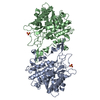 7x0dC 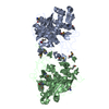 2yijS S: Starting model for refinement C: citing same article ( |
|---|---|
| Similar structure data |
-Links
 PDBj
PDBj- Assembly
| Deposited unit | 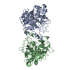
| ||||||||||||
|---|---|---|---|---|---|---|---|---|---|---|---|---|---|
| 1 |
| ||||||||||||
| Unit cell |
|
-Components
| #1: Protein | Mass: 47911.875 Da / Num. of mol.: 2 Source method: isolated from a genetically manipulated source Source: (gene. exp.)  References: UniProt: O49523, Hydrolases; Acting on ester bonds; Carboxylic-ester hydrolases #2: Water | ChemComp-HOH / |  Mass: 18.015 Da / Num. of mol.: 423 / Source method: isolated from a natural source / Formula: H2O Mass: 18.015 Da / Num. of mol.: 423 / Source method: isolated from a natural source / Formula: H2O |
|---|
-Experimental details
-Experiment
| Experiment | Method: X-RAY DIFFRACTION / Number of used crystals: 1 |
|---|
- Sample preparation
| Crystal | Density Matthews: 2.97 Å3/Da / Density % sol: 58.58 % |
|---|---|
| Crystal grow | Temperature: 293 K / Method: vapor diffusion, sitting drop Details: 0.02M calcium chloride, 0.1M sodium acetate, 30% MPD |
-Data collection
| Diffraction | Mean temperature: 93 K / Serial crystal experiment: N |
|---|---|
| Diffraction source | Source: SYNCHROTRON / Site: Photon Factory  / Beamline: BL-17A / Wavelength: 1.0082 Å / Beamline: BL-17A / Wavelength: 1.0082 Å |
| Detector | Type: DECTRIS EIGER X 16M / Detector: PIXEL / Date: Jun 25, 2012 |
| Radiation | Protocol: SINGLE WAVELENGTH / Monochromatic (M) / Laue (L): M / Scattering type: x-ray |
| Radiation wavelength | Wavelength: 1.0082 Å / Relative weight: 1 |
| Reflection | Resolution: 1.799→50 Å / Num. obs: 105892 / % possible obs: 99.8 % / Redundancy: 7.5 % / Biso Wilson estimate: 22.0841058546 Å2 / Rmerge(I) obs: 0.086 / Net I/σ(I): 35.29 |
| Reflection shell | Resolution: 1.8→1.86 Å / Rmerge(I) obs: 0.4 / Mean I/σ(I) obs: 4.7 / Num. unique obs: 4586 |
- Processing
| Software | Name: PHENIX / Version: 1.10.1_2155 / Classification: refinement | |||||||||||||||||||||||||||||||||||||||||||||||||||||||||||||||||||||||||||||||||||||||||||||||||||||||||||||||||||||||||||||||||||||||||||||||||||||||||||||||||||||||||||||||||||||||||||||||||||||||||||||||||||||||||
|---|---|---|---|---|---|---|---|---|---|---|---|---|---|---|---|---|---|---|---|---|---|---|---|---|---|---|---|---|---|---|---|---|---|---|---|---|---|---|---|---|---|---|---|---|---|---|---|---|---|---|---|---|---|---|---|---|---|---|---|---|---|---|---|---|---|---|---|---|---|---|---|---|---|---|---|---|---|---|---|---|---|---|---|---|---|---|---|---|---|---|---|---|---|---|---|---|---|---|---|---|---|---|---|---|---|---|---|---|---|---|---|---|---|---|---|---|---|---|---|---|---|---|---|---|---|---|---|---|---|---|---|---|---|---|---|---|---|---|---|---|---|---|---|---|---|---|---|---|---|---|---|---|---|---|---|---|---|---|---|---|---|---|---|---|---|---|---|---|---|---|---|---|---|---|---|---|---|---|---|---|---|---|---|---|---|---|---|---|---|---|---|---|---|---|---|---|---|---|---|---|---|---|---|---|---|---|---|---|---|---|---|---|---|---|---|---|---|---|
| Refinement | Method to determine structure: MOLECULAR REPLACEMENT Starting model: 2YIJ Resolution: 1.79909514333→33.2867143454 Å / SU ML: 0.167434903764 / Cross valid method: FREE R-VALUE / σ(F): 1.35431739796 / Phase error: 19.6841137382 Stereochemistry target values: GeoStd + Monomer Library + CDL v1.2
| |||||||||||||||||||||||||||||||||||||||||||||||||||||||||||||||||||||||||||||||||||||||||||||||||||||||||||||||||||||||||||||||||||||||||||||||||||||||||||||||||||||||||||||||||||||||||||||||||||||||||||||||||||||||||
| Solvent computation | Shrinkage radii: 0.9 Å / VDW probe radii: 1.11 Å / Solvent model: FLAT BULK SOLVENT MODEL | |||||||||||||||||||||||||||||||||||||||||||||||||||||||||||||||||||||||||||||||||||||||||||||||||||||||||||||||||||||||||||||||||||||||||||||||||||||||||||||||||||||||||||||||||||||||||||||||||||||||||||||||||||||||||
| Displacement parameters | Biso mean: 24.9417230762 Å2 | |||||||||||||||||||||||||||||||||||||||||||||||||||||||||||||||||||||||||||||||||||||||||||||||||||||||||||||||||||||||||||||||||||||||||||||||||||||||||||||||||||||||||||||||||||||||||||||||||||||||||||||||||||||||||
| Refinement step | Cycle: LAST / Resolution: 1.79909514333→33.2867143454 Å
| |||||||||||||||||||||||||||||||||||||||||||||||||||||||||||||||||||||||||||||||||||||||||||||||||||||||||||||||||||||||||||||||||||||||||||||||||||||||||||||||||||||||||||||||||||||||||||||||||||||||||||||||||||||||||
| Refine LS restraints |
| |||||||||||||||||||||||||||||||||||||||||||||||||||||||||||||||||||||||||||||||||||||||||||||||||||||||||||||||||||||||||||||||||||||||||||||||||||||||||||||||||||||||||||||||||||||||||||||||||||||||||||||||||||||||||
| LS refinement shell |
|
