 Movie
Movie Controller

[English] 日本語
 Yorodumi
- PDB-7wzm: Crystal structure of Cytochrome P450 184A1 from streptomyces aver... -
+ Open data

- Basic information
| Entry | Database: PDB / ID: 7wzm | ||||||
|---|---|---|---|---|---|---|---|
| Title | Crystal structure of Cytochrome P450 184A1 from streptomyces avermitilis in complex with Oleic acid | ||||||
 Components Components | Putative cytochrome P450 | ||||||
 Keywords Keywords | OXIDOREDUCTASE / Cytochrome P450 / Streptomyces avermitilis / Oleic acid | ||||||
| Function / homology |  Function and homology information Function and homology informationoxidoreductase activity, acting on paired donors, with incorporation or reduction of molecular oxygen / monooxygenase activity / iron ion binding / heme binding Similarity search - Function | ||||||
| Biological species |  Streptomyces avermitilis (bacteria) Streptomyces avermitilis (bacteria) | ||||||
| Method |  X-RAY DIFFRACTION / SYNCHROTRON / MOLECULAR REPLACEMENT / Resolution: 1.68 Å X-RAY DIFFRACTION / SYNCHROTRON / MOLECULAR REPLACEMENT / Resolution: 1.68 Å | ||||||
 Authors Authors | Kim, V.C. / Kim, D.G. / Lee, S.G. / Lee, G.H. / Lee, S.A. / Kang, L.W. | ||||||
| Funding support | 1items
| ||||||
 Citation Citation | Journal: To Be Published Title: Crystal structure of Cytochrome P450 184A1 from streptomyces avermitilis in complex with Oleic acid Authors: Kim, V.C. / Kim, D.G. / Lee, S.G. / Lee, G.H. / Lee, S.A. / Kang, L.W. | ||||||
| History |
|
- Structure visualization
| Structure viewer | Molecule: MolmilJmol/JSmol |
|---|
- Downloads & links
-Download
| PDBx/mmCIF format | 7wzm.cif.gz | 108.2 KB | Display | PDBx/mmCIF format |
|---|---|---|---|---|
| PDB format | pdb7wzm.ent.gz | 78.3 KB | Display | PDB format |
| PDBx/mmJSON format | 7wzm.json.gz | Tree view | PDBx/mmJSON format | |
| Others |  Other downloads Other downloads |
-Validation report
| Summary document | 7wzm_validation.pdf.gz | 1 MB | Display | wwPDB validaton report |
|---|---|---|---|---|
| Full document | 7wzm_full_validation.pdf.gz | 1.1 MB | Display | |
| Data in XML | 7wzm_validation.xml.gz | 19.9 KB | Display | |
| Data in CIF | 7wzm_validation.cif.gz | 28.7 KB | Display | |
| Arichive directory | https://data.pdbj.org/pub/pdb/validation_reports/wz/7wzmftp://data.pdbj.org/pub/pdb/validation_reports/wz/7wzm | HTTPS FTP |
-Related structure data
| Related structure data |  7wzlS S: Starting model for refinement |
|---|---|
| Similar structure data |
-Links
 PDBj
PDBj


- Assembly
| Deposited unit | 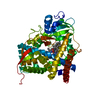
| ||||||||
|---|---|---|---|---|---|---|---|---|---|
| 1 |
| ||||||||
| Unit cell |
|
-Components
| #1: Protein | Mass: 53770.727 Da / Num. of mol.: 1 Source method: isolated from a genetically manipulated source Source: (gene. exp.) Streptomyces avermitilis (bacteria)Strain: ATCC 31267 / DSM 46492 / JCM 5070 / NBRC 14893 / NCIMB 12804 / NRRL 8165 / MA-4680 Gene: cyp21, SAVERM_5111 / Production host: |
|---|---|
| #2: Chemical | ChemComp-OLA /   Mass: 282.461 Da / Num. of mol.: 1 / Source method: obtained synthetically / Formula: C18H34O2 / Feature type: SUBJECT OF INVESTIGATION Mass: 282.461 Da / Num. of mol.: 1 / Source method: obtained synthetically / Formula: C18H34O2 / Feature type: SUBJECT OF INVESTIGATION |
| #3: Chemical | ChemComp-HEM /   Mass: 616.487 Da / Num. of mol.: 1 / Source method: obtained synthetically / Formula: C34H32FeN4O4 Mass: 616.487 Da / Num. of mol.: 1 / Source method: obtained synthetically / Formula: C34H32FeN4O4 |
| #4: Water | ChemComp-HOH /  Mass: 18.015 Da / Num. of mol.: 178 / Source method: isolated from a natural source / Formula: H2O Mass: 18.015 Da / Num. of mol.: 178 / Source method: isolated from a natural source / Formula: H2O |
| Has ligand of interest | Y |
-Experimental details
-Experiment
| Experiment | Method: X-RAY DIFFRACTION / Number of used crystals: 1 |
|---|
- Sample preparation
| Crystal | Density Matthews: 2.11 Å3/Da / Density % sol: 41.61 % |
|---|---|
| Crystal grow | Temperature: 288.15 K / Method: vapor diffusion, sitting drop Details: 15% PEG3350, 0.2 M sodium chloride, 0.1M HEPES, pH 7.0, 5% MPD |
-Data collection
| Diffraction | Mean temperature: 100 K / Serial crystal experiment: N |
|---|---|
| Diffraction source | Source: SYNCHROTRON / Site: PAL/PLS  / Beamline: 5C (4A) / Wavelength: 1 Å / Beamline: 5C (4A) / Wavelength: 1 Å |
| Detector | Type: DECTRIS EIGER2 S 9M / Detector: PIXEL / Date: Dec 2, 2021 |
| Radiation | Protocol: SINGLE WAVELENGTH / Monochromatic (M) / Laue (L): M / Scattering type: x-ray |
| Radiation wavelength | Wavelength: 1 Å / Relative weight: 1 |
| Reflection | Resolution: 1.68→50 Å / Num. obs: 52789 / % possible obs: 99.9 % / Redundancy: 13 % / CC1/2: 0.949 / Net I/σ(I): 36.567 |
| Reflection shell | Resolution: 1.68→1.71 Å / Num. unique obs: 52789 / CC1/2: 0.889 |
- Processing
| Software |
| ||||||||||||||||||||||||||||||||||||||||||||||||||||||||||||
|---|---|---|---|---|---|---|---|---|---|---|---|---|---|---|---|---|---|---|---|---|---|---|---|---|---|---|---|---|---|---|---|---|---|---|---|---|---|---|---|---|---|---|---|---|---|---|---|---|---|---|---|---|---|---|---|---|---|---|---|---|---|
| Refinement | Method to determine structure: MOLECULAR REPLACEMENT Starting model: 7WZL Resolution: 1.68→45.52 Å / Cor.coef. Fo:Fc: 0.947 / Cor.coef. Fo:Fc free: 0.93 / SU B: 1.796 / SU ML: 0.062 / Cross valid method: THROUGHOUT / σ(F): 0 / ESU R: 0.104 / ESU R Free: 0.103 / Stereochemistry target values: MAXIMUM LIKELIHOOD Details: HYDROGENS HAVE BEEN ADDED IN THE RIDING POSITIONS U VALUES : REFINED INDIVIDUALLY
| ||||||||||||||||||||||||||||||||||||||||||||||||||||||||||||
| Solvent computation | Ion probe radii: 0.8 Å / Shrinkage radii: 0.8 Å / VDW probe radii: 1.2 Å / Solvent model: MASK | ||||||||||||||||||||||||||||||||||||||||||||||||||||||||||||
| Displacement parameters | Biso max: 69 Å2 / Biso mean: 14.682 Å2 / Biso min: 6.6 Å2
| ||||||||||||||||||||||||||||||||||||||||||||||||||||||||||||
| Refinement step | Cycle: final / Resolution: 1.68→45.52 Å
| ||||||||||||||||||||||||||||||||||||||||||||||||||||||||||||
| Refine LS restraints |
| ||||||||||||||||||||||||||||||||||||||||||||||||||||||||||||
| LS refinement shell | Resolution: 1.68→1.722 Å / Rfactor Rfree error: 0
|
