 Movie
Movie Controller


+ Open data

- Basic information
| Entry | Database: PDB / ID: 7wzb | ||||||||||||
|---|---|---|---|---|---|---|---|---|---|---|---|---|---|
| Title | lipopolysaccharide assembly protein LapB (open) | ||||||||||||
 Components Components | Lipopolysaccharide assembly protein B | ||||||||||||
 Keywords Keywords | MEMBRANE PROTEIN / lipopolysaccharide assembly protein LapB (open) cytoplasmic soluble domain | ||||||||||||
| Function / homology |  Function and homology information Function and homology informationlipopolysaccharide metabolic process / regulation of lipid biosynthetic process / cytoplasmic side of plasma membrane / transferase activity / iron ion binding Similarity search - Function | ||||||||||||
| Biological species |  Salmonella enterica subsp. enterica serovar Typhimurium str. LT2 (bacteria) Salmonella enterica subsp. enterica serovar Typhimurium str. LT2 (bacteria) | ||||||||||||
| Method |  X-RAY DIFFRACTION / SYNCHROTRON / MOLECULAR REPLACEMENT / Resolution: 2.7 Å X-RAY DIFFRACTION / SYNCHROTRON / MOLECULAR REPLACEMENT / Resolution: 2.7 Å | ||||||||||||
 Authors Authors | Yan, L. / Dong, H. / Li, H. / Liu, X. / Deng, Z. / Dong, C. / Zhang, Z. | ||||||||||||
| Funding support |  China, 3items China, 3items
| ||||||||||||
 Citation Citation | Journal: To Be Published Title: lipopolysaccharide assembly protein LapB (open) Authors: Yan, L. / Dong, H. / Li, H. / Liu, X. / Deng, Z. / Dong, C. / Zhang, Z. | ||||||||||||
| History |
|
- Structure visualization
| Structure viewer | Molecule: MolmilJmol/JSmol |
|---|
- Downloads & links
-Download
| PDBx/mmCIF format | 7wzb.cif.gz | 212.9 KB | Display | PDBx/mmCIF format |
|---|---|---|---|---|
| PDB format | pdb7wzb.ent.gz | 165.9 KB | Display | PDB format |
| PDBx/mmJSON format | 7wzb.json.gz | Tree view | PDBx/mmJSON format | |
| Others |  Other downloads Other downloads |
-Validation report
| Arichive directory | https://data.pdbj.org/pub/pdb/validation_reports/wz/7wzbftp://data.pdbj.org/pub/pdb/validation_reports/wz/7wzb | HTTPS FTP |
|---|
-Related structure data
| Related structure data |  4zlhS S: Starting model for refinement |
|---|---|
| Similar structure data |
-Links
 PDBj
PDBj


- Assembly
| Deposited unit | 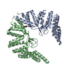
| ||||||||
|---|---|---|---|---|---|---|---|---|---|
| 1 |
| ||||||||
| Unit cell |
|
-Components
| #1: Protein | Mass: 42383.277 Da / Num. of mol.: 2 Source method: isolated from a genetically manipulated source Details: lipopolysaccharide assembly protein B Source: (gene. exp.) Salmonella enterica subsp. enterica serovar Typhimurium str. LT2 (bacteria)Strain: LT2 / Gene: lapB / Plasmid: pet28 / Production host: #2: Chemical |   Mass: 65.409 Da / Num. of mol.: 2 / Source method: obtained synthetically / Formula: Zn Mass: 65.409 Da / Num. of mol.: 2 / Source method: obtained synthetically / Formula: Zn#3: Chemical | ChemComp-PGE / |   Mass: 150.173 Da / Num. of mol.: 1 / Source method: obtained synthetically / Formula: C6H14O4 / Feature type: SUBJECT OF INVESTIGATION Mass: 150.173 Da / Num. of mol.: 1 / Source method: obtained synthetically / Formula: C6H14O4 / Feature type: SUBJECT OF INVESTIGATION#4: Water | ChemComp-HOH / |  Mass: 18.015 Da / Num. of mol.: 17 / Source method: isolated from a natural source / Formula: H2O Mass: 18.015 Da / Num. of mol.: 17 / Source method: isolated from a natural source / Formula: H2OHas ligand of interest | Y | |
|---|
-Experimental details
-Experiment
| Experiment | Method: X-RAY DIFFRACTION / Number of used crystals: 1 |
|---|
- Sample preparation
| Crystal | Density Matthews: 2.08 Å3/Da / Density % sol: 40.92 % |
|---|---|
| Crystal grow | Temperature: 293 K / Method: evaporation / pH: 8 / Details: 21.25% Ethylene glycol 15% glycerol / PH range: 7-8 |
-Data collection
| Diffraction | Mean temperature: 80 K / Serial crystal experiment: N |
|---|---|
| Diffraction source | Source: SYNCHROTRON / Site: Diamond  / Beamline: I03 / Wavelength: 0.9175 Å / Beamline: I03 / Wavelength: 0.9175 Å |
| Detector | Type: DECTRIS PILATUS 2M / Detector: PIXEL / Date: Apr 27, 2015 |
| Radiation | Protocol: SINGLE WAVELENGTH / Monochromatic (M) / Laue (L): M / Scattering type: x-ray |
| Radiation wavelength | Wavelength: 0.9175 Å / Relative weight: 1 |
| Reflection | Resolution: 2.7→24.72 Å / Num. obs: 19835 / % possible obs: 97.03 % / Redundancy: 7.3 % / Biso Wilson estimate: 41.78 Å2 / CC1/2: 1 / CC star: 1 / Rmerge(I) obs: 0.04174 / Rpim(I) all: 0.01674 / Rrim(I) all: 0.04506 / Net I/σ(I): 27.93 |
| Reflection shell | Resolution: 2.7→2.796 Å / Redundancy: 7.08 % / Rmerge(I) obs: 0.2306 / Mean I/σ(I) obs: 7.08 / Num. unique obs: 1537 / CC1/2: 0.991 / CC star: 0.998 / Rpim(I) all: 0.08943 / Rrim(I) all: 0.2476 / % possible all: 78.14 |
- Processing
| Software |
| ||||||||||||||||||||||||||||||||||||||||
|---|---|---|---|---|---|---|---|---|---|---|---|---|---|---|---|---|---|---|---|---|---|---|---|---|---|---|---|---|---|---|---|---|---|---|---|---|---|---|---|---|---|
| Refinement | Method to determine structure: MOLECULAR REPLACEMENT Starting model: 4ZLH Resolution: 2.7→24.46 Å / Cross valid method: THROUGHOUT / Stereochemistry target values: ML
| ||||||||||||||||||||||||||||||||||||||||
| Solvent computation | Solvent model: FLAT BULK SOLVENT MODEL | ||||||||||||||||||||||||||||||||||||||||
| Displacement parameters | Biso max: 148.98 Å2 / Biso mean: 73.9629 Å2 / Biso min: 37.84 Å2 | ||||||||||||||||||||||||||||||||||||||||
| Refinement step | Cycle: final / Resolution: 2.7→24.46 Å
| ||||||||||||||||||||||||||||||||||||||||
| LS refinement shell | Resolution: 2.7→2.796 Å / Rfactor Rfree error: 0
| ||||||||||||||||||||||||||||||||||||||||
| Refinement TLS params. | Method: refined / Origin x: 13.8983 Å / Origin y: -17.2672 Å / Origin z: -9.0542 Å
| ||||||||||||||||||||||||||||||||||||||||
| Refinement TLS group |
|
