 ムービー
ムービー コントローラー


+ データを開く

- 基本情報
基本情報
| 登録情報 | データベース: PDB / ID: 7wz8 | ||||||
|---|---|---|---|---|---|---|---|
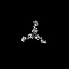
| タイトル | Structure of human langerin complex in Birbeck granules | ||||||
 要素 要素 | SNAP-tag,C-type lectin domain family 4 member K | ||||||
 キーワード キーワード | MEMBRANE PROTEIN / Birbeck granule / C-type lectin / Langerhans cell / virus defense | ||||||
| 機能・相同性 |  機能・相同性情報 機能・相同性情報Cross-presentation of soluble exogenous antigens (endosomes) / pattern recognition receptor activity / D-mannose binding / endocytic vesicle / clathrin-coated endocytic vesicle membrane / carbohydrate binding / early endosome membrane / defense response to virus / immune response / external side of plasma membrane / plasma membrane 類似検索 - 分子機能 | ||||||
| 生物種 |  Homo sapiens (ヒト) Homo sapiens (ヒト) | ||||||
| 手法 | 電子顕微鏡法 / サブトモグラム平均法 / クライオ電子顕微鏡法 / 解像度: 6.4 Å | ||||||
 データ登録者 データ登録者 | Oda, T. / Yanagisawa, H. | ||||||
| 資金援助 |  日本, 1件 日本, 1件
| ||||||
 引用 引用 | ジャーナル: Elife / 年: 2022 タイトル: Cryo-electron tomography of Birbeck granules reveals the molecular mechanism of langerin lattice formation. 著者: Toshiyuki Oda / Haruaki Yanagisawa / Hideyuki Shinmori / Youichi Ogawa / Tatsuyoshi Kawamura / 要旨: Langerhans cells are specialized antigen-presenting cells localized within the epidermis and mucosal epithelium. Upon contact with Langerhans cells, pathogens are captured by the C-type lectin ...Langerhans cells are specialized antigen-presenting cells localized within the epidermis and mucosal epithelium. Upon contact with Langerhans cells, pathogens are captured by the C-type lectin langerin and internalized into a structurally unique vesicle known as a Birbeck granule. Although the immunological role of Langerhans cells and Birbeck granules have been extensively studied, the mechanism by which the characteristic zippered membrane structure of Birbeck granules is formed remains elusive. In this study, we observed isolated Birbeck granules using cryo-electron tomography and reconstructed the 3D structure of the repeating unit of the honeycomb lattice of langerin at 6.4 Å resolution. We found that the interaction between the two langerin trimers was mediated by docking the flexible loop at residues 258-263 into the secondary carbohydrate-binding cleft. Mutations within the loop inhibited Birbeck granule formation and the internalization of HIV pseudovirus. These findings suggest a molecular mechanism for membrane zippering during Birbeck granule biogenesis and provide insight into the role of langerin in the defense against viral infection. | ||||||
| 履歴 |
|
- 構造の表示
構造の表示
| 構造ビューア | 分子: MolmilJmol/JSmol |
|---|
- ダウンロードとリンク
-ダウンロード
| PDBx/mmCIF形式 | 7wz8.cif.gz | 292.4 KB | 表示 | PDBx/mmCIF形式 |
|---|---|---|---|---|
| PDB形式 | pdb7wz8.ent.gz | 表示 | PDB形式 | |
| PDBx/mmJSON形式 | 7wz8.json.gz | ツリー表示 | PDBx/mmJSON形式 | |
| その他 |  その他のダウンロード その他のダウンロード |
-検証レポート
| 文書・要旨 | 7wz8_validation.pdf.gz | 947.9 KB | 表示 | wwPDB検証レポート |
|---|---|---|---|---|
| 文書・詳細版 | 7wz8_full_validation.pdf.gz | 973.8 KB | 表示 | |
| XML形式データ | 7wz8_validation.xml.gz | 40.5 KB | 表示 | |
| CIF形式データ | 7wz8_validation.cif.gz | 60.8 KB | 表示 | |
| アーカイブディレクトリ | https://data.pdbj.org/pub/pdb/validation_reports/wz/7wz8ftp://data.pdbj.org/pub/pdb/validation_reports/wz/7wz8 | HTTPS FTP |
-関連構造データ
-リンク
 PDBj
PDBj


- 集合体
集合体
| 登録構造単位 | 
|
|---|---|
| 1 |
|
-要素
| #1: タンパク質 | 分子量: 60547.773 Da / 分子数: 6 / 由来タイプ: 組換発現 詳細: 185 amino acids SNAP-tag + 3xHA tag (YPYDVPDYAYPYDVPDYAYPYDVPDYA) + Linker (GSSG) + HRV3C cleavage site (LEVLFQGP) 由来: (組換発現) Homo sapiens (ヒト) / 遺伝子: CD207, CLEC4K / 細胞株 (発現宿主): 293T / 発現宿主: Homo sapiens (ヒト) / 参照: UniProt: Q9UJ71Has protein modification | Y | |
|---|
-実験情報
-実験
| 実験 | 手法: 電子顕微鏡法 |
|---|---|
| EM実験 | 試料の集合状態: 2D ARRAY / 3次元再構成法: サブトモグラム平均法 |
- 試料調製
試料調製
| 構成要素 | 名称: Repeating unit of langerin lattice in Birbeck granule タイプ: COMPLEX 詳細: Birbeck granules isolated from 293T cells by immunoprecipitation. Entity ID: all / 由来: RECOMBINANT |
|---|---|
| 分子量 | 実験値: NO |
| 由来(天然) | 生物種: Homo sapiens (ヒト) |
| 由来(組換発現) | 生物種: Homo sapiens (ヒト) / 細胞: 293T / プラスミド: pcDNA3.1 |
| 緩衝液 | pH: 7.2 |
| 試料 | 濃度: 0.05 mg/ml / 包埋: NO / シャドウイング: NO / 染色: NO / 凍結: YES |
| 試料支持 | グリッドの材料: COPPER/RHODIUM / グリッドのサイズ: 200 divisions/in. / グリッドのタイプ: Quantifoil R1.2/1.3 |
| 急速凍結 | 装置: FEI VITROBOT MARK IV / 凍結剤: ETHANE / 湿度: 100 % / 凍結前の試料温度: 277 K / 詳細: Blot for 10 seconds before plunging |
- 電子顕微鏡撮影
| 実験機器 |  モデル: Titan Krios / 画像提供: FEI Company |
|---|---|
| 顕微鏡 | モデル: TFS KRIOS |
| 電子銃 | 電子線源:  FIELD EMISSION GUN / 加速電圧: 300 kV / 照射モード: FLOOD BEAM FIELD EMISSION GUN / 加速電圧: 300 kV / 照射モード: FLOOD BEAM |
| 電子レンズ | モード: BRIGHT FIELD / 倍率(公称値): 35000 X / 最大 デフォーカス(公称値): 6000 nm / 最小 デフォーカス(公称値): 3000 nm / Calibrated defocus min: 2600 nm / 最大 デフォーカス(補正後): 8500 nm / Cs: 2.7 mm |
| 試料ホルダ | 凍結剤: NITROGEN 試料ホルダーモデル: FEI TITAN KRIOS AUTOGRID HOLDER |
| 撮影 | 平均露光時間: 0.74 sec. / 電子線照射量: 1.24 e/Å2 / Avg electron dose per subtomogram: 49.6 e/Å2 / フィルム・検出器のモデル: GATAN K3 (6k x 4k) / 撮影したグリッド数: 1 |
| 電子光学装置 | エネルギーフィルター名称: GIF Quantum LS / エネルギーフィルタースリット幅: 20 eV |
- 解析
解析
| EMソフトウェア |
| ||||||||||||||||||||||||||||||||||||||||
|---|---|---|---|---|---|---|---|---|---|---|---|---|---|---|---|---|---|---|---|---|---|---|---|---|---|---|---|---|---|---|---|---|---|---|---|---|---|---|---|---|---|
| CTF補正 | タイプ: PHASE FLIPPING AND AMPLITUDE CORRECTION | ||||||||||||||||||||||||||||||||||||||||
| 対称性 | 点対称性: C3 (3回回転対称) | ||||||||||||||||||||||||||||||||||||||||
| 3次元再構成 | 解像度: 6.4 Å / 解像度の算出法: FSC 0.143 CUT-OFF / 粒子像の数: 63563 / クラス平均像の数: 1 / 対称性のタイプ: POINT | ||||||||||||||||||||||||||||||||||||||||
| EM volume selection | Num. of tomograms: 33 / Num. of volumes extracted: 93953 | ||||||||||||||||||||||||||||||||||||||||
| 原子モデル構築 | プロトコル: BACKBONE TRACE | ||||||||||||||||||||||||||||||||||||||||
| 原子モデル構築 | PDB-ID: 3KQG Accession code: 3KQG / Source name: PDB / タイプ: experimental model |

