 Movie
Movie Controller

[English] 日本語
 Yorodumi
- PDB-7wyr: Crystal structure of Cypovirus Polyhedra mutant fused with CLN025 -
+ Open data

- Basic information
| Entry | Database: PDB / ID: 7wyr | ||||||
|---|---|---|---|---|---|---|---|
| Title | Crystal structure of Cypovirus Polyhedra mutant fused with CLN025 | ||||||
 Components Components | Polyhedrin fused with CLN025 | ||||||
 Keywords Keywords | VIRAL PROTEIN | ||||||
| Function / homology | Cypovirus polyhedrin, Cypovirus 1 type / Cypovirus polyhedrin protein / viral occlusion body / host cell cytoplasm / Polyhedrin Function and homology information Function and homology information | ||||||
| Biological species |   Bombyx mori cypovirus 1 Bombyx mori cypovirus 1synthetic construct (others) | ||||||
| Method |  X-RAY DIFFRACTION / SYNCHROTRON / MOLECULAR REPLACEMENT / Resolution: 1.75 Å X-RAY DIFFRACTION / SYNCHROTRON / MOLECULAR REPLACEMENT / Resolution: 1.75 Å | ||||||
 Authors Authors | Kojima, M. / Abe, S. / Hirata, K. / Yamashita, K. / Ueno, T. | ||||||
| Funding support |  Japan, 1items Japan, 1items
| ||||||
 Citation Citation | Journal: Biomater Sci / Year: 2023 Title: Engineering of an in-cell protein crystal for fastening a metastable conformation of a target miniprotein. Authors: Kojima, M. / Abe, S. / Furuta, T. / Tran, D.P. / Hirata, K. / Yamashita, K. / Hishikawa, Y. / Kitao, A. / Ueno, T. | ||||||
| History |
|
- Structure visualization
| Structure viewer | Molecule: MolmilJmol/JSmol |
|---|
- Downloads & links
-Download
| PDBx/mmCIF format | 7wyr.cif.gz | 70.3 KB | Display | PDBx/mmCIF format |
|---|---|---|---|---|
| PDB format | pdb7wyr.ent.gz | 48 KB | Display | PDB format |
| PDBx/mmJSON format | 7wyr.json.gz | Tree view | PDBx/mmJSON format | |
| Others |  Other downloads Other downloads |
-Validation report
| Arichive directory | https://data.pdbj.org/pub/pdb/validation_reports/wy/7wyrftp://data.pdbj.org/pub/pdb/validation_reports/wy/7wyr | HTTPS FTP |
|---|
-Related structure data
| Related structure data | 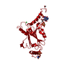 2oh6S S: Starting model for refinement |
|---|---|
| Similar structure data |
-Links
 PDBj
PDBj- Assembly
| Deposited unit | 
| ||||||||
|---|---|---|---|---|---|---|---|---|---|
| 1 |
| ||||||||
| Unit cell |
| ||||||||
| Components on special symmetry positions |
|
-Components
| #1: Protein | Mass: 29053.986 Da / Num. of mol.: 1 / Fragment: UNP residues 2-71, UNP residues 77-248 Source method: isolated from a genetically manipulated source Source: (gene. exp.) Bombyx mori cypovirus 1, (gene. exp.) synthetic construct (others)Production host:   Spodoptera frugiperda (fall armyworm) / References: UniProt: P11041 Spodoptera frugiperda (fall armyworm) / References: UniProt: P11041 | ||||||
|---|---|---|---|---|---|---|---|
| #2: Chemical | ChemComp-CL /   Mass: 35.453 Da / Num. of mol.: 1 / Source method: obtained synthetically / Formula: Cl / Feature type: SUBJECT OF INVESTIGATION Mass: 35.453 Da / Num. of mol.: 1 / Source method: obtained synthetically / Formula: Cl / Feature type: SUBJECT OF INVESTIGATION | ||||||
| #3: Chemical |   Mass: 62.068 Da / Num. of mol.: 2 / Source method: obtained synthetically / Formula: C2H6O2 / Feature type: SUBJECT OF INVESTIGATION Mass: 62.068 Da / Num. of mol.: 2 / Source method: obtained synthetically / Formula: C2H6O2 / Feature type: SUBJECT OF INVESTIGATION#4: Water | ChemComp-HOH / |  Mass: 18.015 Da / Num. of mol.: 118 / Source method: isolated from a natural source / Formula: H2O Mass: 18.015 Da / Num. of mol.: 118 / Source method: isolated from a natural source / Formula: H2OHas ligand of interest | Y | Has protein modification | Y | |
-Experimental details
-Experiment
| Experiment | Method: X-RAY DIFFRACTION / Number of used crystals: 1 |
|---|
- Sample preparation
| Crystal | Density Matthews: 1.65 Å3/Da / Density % sol: 25.25 % |
|---|---|
| Crystal grow | Temperature: 293 K / Method: in cell / Details: in cell crystallization |
-Data collection
| Diffraction | Mean temperature: 100 K / Serial crystal experiment: N |
|---|---|
| Diffraction source | Source: SYNCHROTRON / Site: SPring-8 / Beamline: BL32XU / Wavelength: 1 Å |
| Detector | Type: DECTRIS PILATUS3 6M / Detector: PIXEL / Date: Dec 5, 2019 |
| Radiation | Monochromator: M / Protocol: SINGLE WAVELENGTH / Monochromatic (M) / Laue (L): M / Scattering type: x-ray |
| Radiation wavelength | Wavelength: 1 Å / Relative weight: 1 |
| Reflection | Resolution: 1.75→50 Å / Num. obs: 19331 / % possible obs: 100 % / Redundancy: 211.2 % / CC1/2: 0.98 / Net I/σ(I): 5.84 |
| Reflection shell | Resolution: 1.75→1.76 Å / Num. unique obs: 489 / CC1/2: 0.319 |
- Processing
| Software |
| ||||||||||||||||||||||||||||||||||||||||||||||||||||||||||||||||||||||||||||||||||||||||||||||||||||||||||||||||||||||||||||||||||||||||||||||||||||||
|---|---|---|---|---|---|---|---|---|---|---|---|---|---|---|---|---|---|---|---|---|---|---|---|---|---|---|---|---|---|---|---|---|---|---|---|---|---|---|---|---|---|---|---|---|---|---|---|---|---|---|---|---|---|---|---|---|---|---|---|---|---|---|---|---|---|---|---|---|---|---|---|---|---|---|---|---|---|---|---|---|---|---|---|---|---|---|---|---|---|---|---|---|---|---|---|---|---|---|---|---|---|---|---|---|---|---|---|---|---|---|---|---|---|---|---|---|---|---|---|---|---|---|---|---|---|---|---|---|---|---|---|---|---|---|---|---|---|---|---|---|---|---|---|---|---|---|---|---|---|---|---|
| Refinement | Method to determine structure: MOLECULAR REPLACEMENT Starting model: 2OH6 Resolution: 1.75→42.707 Å / Cor.coef. Fo:Fc: 0.947 / Cor.coef. Fo:Fc free: 0.922 / SU B: 2.799 / SU ML: 0.085 / Cross valid method: FREE R-VALUE / ESU R: 0.157 / ESU R Free: 0.134
| ||||||||||||||||||||||||||||||||||||||||||||||||||||||||||||||||||||||||||||||||||||||||||||||||||||||||||||||||||||||||||||||||||||||||||||||||||||||
| Solvent computation | Ion probe radii: 0.8 Å / Shrinkage radii: 0.8 Å / VDW probe radii: 1.2 Å / Solvent model: MASK BULK SOLVENT | ||||||||||||||||||||||||||||||||||||||||||||||||||||||||||||||||||||||||||||||||||||||||||||||||||||||||||||||||||||||||||||||||||||||||||||||||||||||
| Displacement parameters | Biso mean: 9.137 Å2
| ||||||||||||||||||||||||||||||||||||||||||||||||||||||||||||||||||||||||||||||||||||||||||||||||||||||||||||||||||||||||||||||||||||||||||||||||||||||
| Refinement step | Cycle: LAST / Resolution: 1.75→42.707 Å
| ||||||||||||||||||||||||||||||||||||||||||||||||||||||||||||||||||||||||||||||||||||||||||||||||||||||||||||||||||||||||||||||||||||||||||||||||||||||
| Refine LS restraints |
| ||||||||||||||||||||||||||||||||||||||||||||||||||||||||||||||||||||||||||||||||||||||||||||||||||||||||||||||||||||||||||||||||||||||||||||||||||||||
| LS refinement shell |
|
