 Movie
Movie Controller

[English] 日本語
 Yorodumi
- PDB-7wwx: Crystal structure of Herbaspirillum huttiense L-arabinose 1-dehyd... -
+ Open data

- Basic information
| Entry | Database: PDB / ID: 7wwx | ||||||
|---|---|---|---|---|---|---|---|
| Title | Crystal structure of Herbaspirillum huttiense L-arabinose 1-dehydrogenase (NAD bound form) | ||||||
 Components Components | NAD(P)-dependent dehydrogenase (Short-subunit alcohol dehydrogenase family) | ||||||
 Keywords Keywords | OXIDOREDUCTASE / L-ARABINOSE METABOLISM / NAD-DEPENDENT DEHYDROGENASE / SDR PROTEIN FAMILY | ||||||
| Function / homology | NICOTINAMIDE-ADENINE-DINUCLEOTIDE / DI(HYDROXYETHYL)ETHER / :  Function and homology information Function and homology information | ||||||
| Biological species |  Herbaspirillum huttiense subsp. putei IAM 15032 (bacteria) Herbaspirillum huttiense subsp. putei IAM 15032 (bacteria) | ||||||
| Method |  X-RAY DIFFRACTION / SYNCHROTRON / MOLECULAR REPLACEMENT / Resolution: 1.36 Å X-RAY DIFFRACTION / SYNCHROTRON / MOLECULAR REPLACEMENT / Resolution: 1.36 Å | ||||||
 Authors Authors | Matsubara, R. / Yoshiwara, K. / Watanabe, Y. / Watanabe, S. | ||||||
| Funding support | 1items
| ||||||
 Citation Citation | Journal: Biochem.Biophys.Res.Commun. / Year: 2022 Title: Crystal structure of L-arabinose 1-dehydrogenase as a short-chain reductase/dehydrogenase protein. Authors: Watanabe, S. / Yoshiwara, K. / Matsubara, R. / Watanabe, Y. | ||||||
| History |
|
- Structure visualization
| Structure viewer | Molecule: MolmilJmol/JSmol |
|---|
- Downloads & links
-Download
| PDBx/mmCIF format | 7wwx.cif.gz | 125.7 KB | Display | PDBx/mmCIF format |
|---|---|---|---|---|
| PDB format | pdb7wwx.ent.gz | 94.4 KB | Display | PDB format |
| PDBx/mmJSON format | 7wwx.json.gz | Tree view | PDBx/mmJSON format | |
| Others |  Other downloads Other downloads |
-Validation report
| Arichive directory | https://data.pdbj.org/pub/pdb/validation_reports/ww/7wwxftp://data.pdbj.org/pub/pdb/validation_reports/ww/7wwx | HTTPS FTP |
|---|
-Related structure data
| Related structure data | 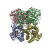 3nugS S: Starting model for refinement |
|---|---|
| Similar structure data |
-Links
 PDBj
PDBj- Assembly
| Deposited unit | 
| ||||||||
|---|---|---|---|---|---|---|---|---|---|
| 1 | 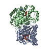
| ||||||||
| Unit cell |
|
-Components
| #1: Protein | Mass: 29717.631 Da / Num. of mol.: 2 Source method: isolated from a genetically manipulated source Source: (gene. exp.) Herbaspirillum huttiense subsp. putei IAM 15032 (bacteria)Gene: DFS02_4196 / Production host: #2: Chemical |   Mass: 663.425 Da / Num. of mol.: 2 / Source method: obtained synthetically / Formula: C21H27N7O14P2 / Feature type: SUBJECT OF INVESTIGATION / Comment: NAD*YM Mass: 663.425 Da / Num. of mol.: 2 / Source method: obtained synthetically / Formula: C21H27N7O14P2 / Feature type: SUBJECT OF INVESTIGATION / Comment: NAD*YM#3: Chemical | ChemComp-PEG / |   Mass: 106.120 Da / Num. of mol.: 1 / Source method: obtained synthetically / Formula: C4H10O3 Mass: 106.120 Da / Num. of mol.: 1 / Source method: obtained synthetically / Formula: C4H10O3#4: Water | ChemComp-HOH / |  Mass: 18.015 Da / Num. of mol.: 550 / Source method: isolated from a natural source / Formula: H2O Mass: 18.015 Da / Num. of mol.: 550 / Source method: isolated from a natural source / Formula: H2OHas ligand of interest | Y | |
|---|
-Experimental details
-Experiment
| Experiment | Method: X-RAY DIFFRACTION / Number of used crystals: 1 |
|---|
- Sample preparation
| Crystal | Density Matthews: 2.29 Å3/Da / Density % sol: 46.34 % |
|---|---|
| Crystal grow | Temperature: 293 K / Method: vapor diffusion, sitting drop / Details: 0.2M Sodium formate, 20% PEG3350 |
-Data collection
| Diffraction | Mean temperature: 100 K / Serial crystal experiment: N | ||||||||||||||||||||||||||||||
|---|---|---|---|---|---|---|---|---|---|---|---|---|---|---|---|---|---|---|---|---|---|---|---|---|---|---|---|---|---|---|---|
| Diffraction source | Source: SYNCHROTRON / Site: SPring-8  / Beamline: BL45XU / Wavelength: 1 Å / Beamline: BL45XU / Wavelength: 1 Å | ||||||||||||||||||||||||||||||
| Detector | Type: DECTRIS PILATUS 6M / Detector: PIXEL / Date: Oct 21, 2021 | ||||||||||||||||||||||||||||||
| Radiation | Protocol: SINGLE WAVELENGTH / Monochromatic (M) / Laue (L): M / Scattering type: x-ray | ||||||||||||||||||||||||||||||
| Radiation wavelength | Wavelength: 1 Å / Relative weight: 1 | ||||||||||||||||||||||||||||||
| Reflection | Resolution: 1.36→42.82 Å / Num. obs: 114656 / % possible obs: 99.8 % / Redundancy: 9.9 % / CC1/2: 0.993 / Rmerge(I) obs: 0.236 / Rpim(I) all: 0.079 / Rrim(I) all: 0.249 / Net I/σ(I): 8.3 | ||||||||||||||||||||||||||||||
| Reflection shell | Diffraction-ID: 1
|
- Processing
| Software |
| ||||||||||||||||||||||||||||||||||||||||||||||||||||||||||||
|---|---|---|---|---|---|---|---|---|---|---|---|---|---|---|---|---|---|---|---|---|---|---|---|---|---|---|---|---|---|---|---|---|---|---|---|---|---|---|---|---|---|---|---|---|---|---|---|---|---|---|---|---|---|---|---|---|---|---|---|---|---|
| Refinement | Method to determine structure: MOLECULAR REPLACEMENT Starting model: 3NUG Resolution: 1.36→42.82 Å / Cor.coef. Fo:Fc: 0.967 / Cor.coef. Fo:Fc free: 0.958 / SU B: 0.979 / SU ML: 0.039 / Cross valid method: THROUGHOUT / σ(F): 0 / ESU R: 0.052 / ESU R Free: 0.054 / Stereochemistry target values: MAXIMUM LIKELIHOOD Details: HYDROGENS HAVE BEEN ADDED IN THE RIDING POSITIONS U VALUES : REFINED INDIVIDUALLY
| ||||||||||||||||||||||||||||||||||||||||||||||||||||||||||||
| Solvent computation | Ion probe radii: 0.8 Å / Shrinkage radii: 0.8 Å / VDW probe radii: 1.2 Å / Solvent model: MASK | ||||||||||||||||||||||||||||||||||||||||||||||||||||||||||||
| Displacement parameters | Biso max: 51.82 Å2 / Biso mean: 12.04 Å2 / Biso min: 5.65 Å2
| ||||||||||||||||||||||||||||||||||||||||||||||||||||||||||||
| Refinement step | Cycle: final / Resolution: 1.36→42.82 Å
| ||||||||||||||||||||||||||||||||||||||||||||||||||||||||||||
| Refine LS restraints |
| ||||||||||||||||||||||||||||||||||||||||||||||||||||||||||||
| LS refinement shell | Resolution: 1.36→1.395 Å / Rfactor Rfree error: 0 / Total num. of bins used: 20
|
