 Movie
Movie Controller


+ Open data

- Basic information
| Entry | Database: PDB / ID: 7ww3 | ||||||
|---|---|---|---|---|---|---|---|
| Title | Crystal structure of MmIMP1-KH34 tandem domain | ||||||
 Components Components | Insulin-like growth factor 2 mRNA-binding protein 1 | ||||||
 Keywords Keywords | RNA BINDING PROTEIN / IMP1 / IGF2BP1 / ZBP1 / RNA binding / RBP / splicing | ||||||
| Function / homology |  Function and homology information Function and homology informationregulation of mRNA stability involved in response to stress / pallium cell proliferation in forebrain / CRD-mediated mRNA stability complex / RNA localization / negative regulation of nuclear-transcribed mRNA catabolic process, deadenylation-dependent decay / CRD-mediated mRNA stabilization / dendrite arborization / neuronal stem cell population maintenance / N6-methyladenosine-containing RNA reader activity / positive regulation of cytoplasmic translation ...regulation of mRNA stability involved in response to stress / pallium cell proliferation in forebrain / CRD-mediated mRNA stability complex / RNA localization / negative regulation of nuclear-transcribed mRNA catabolic process, deadenylation-dependent decay / CRD-mediated mRNA stabilization / dendrite arborization / neuronal stem cell population maintenance / N6-methyladenosine-containing RNA reader activity / positive regulation of cytoplasmic translation / mRNA transport / translation regulator activity / mRNA 3'-UTR binding / filopodium / P-body / mRNA 5'-UTR binding / cytoplasmic stress granule / nervous system development / lamellipodium / growth cone / dendritic spine / negative regulation of translation / ribonucleoprotein complex / axon / neuronal cell body / mRNA binding / dendrite / perinuclear region of cytoplasm / nucleoplasm / nucleus / cytoplasm / cytosol Similarity search - Function | ||||||
| Biological species |  | ||||||
| Method |  X-RAY DIFFRACTION / SYNCHROTRON / MOLECULAR REPLACEMENT / Resolution: 1.9 Å X-RAY DIFFRACTION / SYNCHROTRON / MOLECULAR REPLACEMENT / Resolution: 1.9 Å | ||||||
 Authors Authors | Li, X.J. / Wu, B.X. | ||||||
| Funding support | 1items
| ||||||
 Citation Citation | Journal: Structure / Year: 2025 Title: Structural basis for the RNA binding properties of mouse IGF2BP3. Authors: Li, X. / Guo, W. / Wen, Y. / Meng, C. / Zhang, Q. / Chen, H. / Zhao, X. / Wu, B. | ||||||
| History |
|
- Structure visualization
| Structure viewer | Molecule: MolmilJmol/JSmol |
|---|
- Downloads & links
-Download
| PDBx/mmCIF format | 7ww3.cif.gz | 91.7 KB | Display | PDBx/mmCIF format |
|---|---|---|---|---|
| PDB format | pdb7ww3.ent.gz | 58.1 KB | Display | PDB format |
| PDBx/mmJSON format | 7ww3.json.gz | Tree view | PDBx/mmJSON format | |
| Others |  Other downloads Other downloads |
-Validation report
| Arichive directory | https://data.pdbj.org/pub/pdb/validation_reports/ww/7ww3ftp://data.pdbj.org/pub/pdb/validation_reports/ww/7ww3 | HTTPS FTP |
|---|
-Related structure data
| Related structure data |  7vklC  7vsjC  7yewC  7yexC  7yeyC 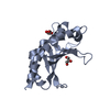 3krmS S: Starting model for refinement C: citing same article ( |
|---|---|
| Similar structure data |
-Links
 PDBj
PDBj
- Assembly
| Deposited unit | 
| ||||||||||||
|---|---|---|---|---|---|---|---|---|---|---|---|---|---|
| 1 |
| ||||||||||||
| Unit cell |
|
-Components
| #1: Protein | Mass: 19654.705 Da / Num. of mol.: 1 Source method: isolated from a genetically manipulated source Source: (gene. exp.)  |
|---|---|
| #2: Water | ChemComp-HOH /  Mass: 18.015 Da / Num. of mol.: 40 / Source method: isolated from a natural source / Formula: H2O Mass: 18.015 Da / Num. of mol.: 40 / Source method: isolated from a natural source / Formula: H2O |
| Has protein modification | N |
-Experimental details
-Experiment
| Experiment | Method: X-RAY DIFFRACTION / Number of used crystals: 1 |
|---|
- Sample preparation
| Crystal | Density Matthews: 2.45 Å3/Da / Density % sol: 49.71 % |
|---|---|
| Crystal grow | Temperature: 293 K / Method: vapor diffusion, hanging drop / pH: 8.5 Details: 0.2 M Magnesium chloride hexahydrate, 0.1 M TRIS hydrochloride pH 8.5, 30% w/v Polyethylene glycol 4000 |
-Data collection
| Diffraction | Mean temperature: 100 K / Serial crystal experiment: N |
|---|---|
| Diffraction source | Source: SYNCHROTRON / Site: SSRF  / Beamline: BL19U1 / Wavelength: 0.97852 Å / Beamline: BL19U1 / Wavelength: 0.97852 Å |
| Detector | Type: DECTRIS PILATUS 6M / Detector: PIXEL / Date: Jan 21, 2022 |
| Radiation | Protocol: SINGLE WAVELENGTH / Monochromatic (M) / Laue (L): M / Scattering type: x-ray |
| Radiation wavelength | Wavelength: 0.97852 Å / Relative weight: 1 |
| Reflection | Resolution: 1.9→30 Å / Num. obs: 16426 / % possible obs: 99.8 % / Redundancy: 37 % / Biso Wilson estimate: 42.01 Å2 / CC1/2: 1 / Rmerge(I) obs: 0.075 / Rpim(I) all: 0.012 / Rrim(I) all: 0.076 / Net I/σ(I): 30.2 |
| Reflection shell | Resolution: 1.9→2 Å / Redundancy: 37.6 % / Rmerge(I) obs: 1.471 / Mean I/σ(I) obs: 3 / Num. unique obs: 2311 / CC1/2: 0.869 / Rpim(I) all: 0.237 / Rrim(I) all: 1.49 / % possible all: 99.6 |
- Processing
| Software |
| |||||||||||||||||||||||||||||||||||||||||||||||||
|---|---|---|---|---|---|---|---|---|---|---|---|---|---|---|---|---|---|---|---|---|---|---|---|---|---|---|---|---|---|---|---|---|---|---|---|---|---|---|---|---|---|---|---|---|---|---|---|---|---|---|
| Refinement | Method to determine structure: MOLECULAR REPLACEMENT Starting model: 3krm Resolution: 1.9→26.84 Å / SU ML: 0.1935 / Cross valid method: FREE R-VALUE / σ(F): 1.34 / Phase error: 30.9658 Stereochemistry target values: GeoStd + Monomer Library + CDL v1.2
| |||||||||||||||||||||||||||||||||||||||||||||||||
| Solvent computation | Shrinkage radii: 0.9 Å / VDW probe radii: 1.11 Å / Solvent model: FLAT BULK SOLVENT MODEL | |||||||||||||||||||||||||||||||||||||||||||||||||
| Displacement parameters | Biso mean: 54.55 Å2 | |||||||||||||||||||||||||||||||||||||||||||||||||
| Refinement step | Cycle: LAST / Resolution: 1.9→26.84 Å
| |||||||||||||||||||||||||||||||||||||||||||||||||
| Refine LS restraints |
| |||||||||||||||||||||||||||||||||||||||||||||||||
| LS refinement shell |
| |||||||||||||||||||||||||||||||||||||||||||||||||
| Refinement TLS params. | Method: refined / Origin x: 29.6090431825 Å / Origin y: -12.4275685093 Å / Origin z: -3.13449606709 Å
| |||||||||||||||||||||||||||||||||||||||||||||||||
| Refinement TLS group | Selection details: all |
