 Movie
Movie Controller

[English] 日本語
 Yorodumi
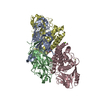
- PDB-7wux: Crystal structure of AziU3/U2 complexed with (5S,6S)-O7-sulfo DAD... -
+ Open data

- Basic information
| Entry | Database: PDB / ID: 7wux | ||||||||||||
|---|---|---|---|---|---|---|---|---|---|---|---|---|---|
| Title | Crystal structure of AziU3/U2 complexed with (5S,6S)-O7-sulfo DADH from Streptomyces sahachiroi | ||||||||||||
 Components Components |
| ||||||||||||
 Keywords Keywords | BIOSYNTHETIC PROTEIN / ES Complex / New Fold / Aziridine synthase / Azinomycin B | ||||||||||||
| Function / homology | Butirosin biosynthesis protein H, N-terminal / Butirosin biosynthesis protein H, N-terminal / Chem-6OI / 1-ETHOXY-2-(2-ETHOXYETHOXY)ETHANE / TRIETHYLENE GLYCOL / Azi28 / Azi29 Function and homology information Function and homology information | ||||||||||||
| Biological species |  Streptomyces sahachiroi (bacteria) Streptomyces sahachiroi (bacteria) | ||||||||||||
| Method |  X-RAY DIFFRACTION / SYNCHROTRON / SAD / Resolution: 1.8 Å X-RAY DIFFRACTION / SYNCHROTRON / SAD / Resolution: 1.8 Å | ||||||||||||
 Authors Authors | Kurosawa, S. / Yoshida, A. / Tomita, T. / Nishiyama, M. | ||||||||||||
| Funding support |  Japan, 3items Japan, 3items
| ||||||||||||
 Citation Citation | Journal: J.Am.Chem.Soc. / Year: 2022 Title: Molecular Basis for Enzymatic Aziridine Formation via Sulfate Elimination. Authors: Kurosawa, S. / Hasebe, F. / Okamura, H. / Yoshida, A. / Matsuda, K. / Sone, Y. / Tomita, T. / Shinada, T. / Takikawa, H. / Kuzuyama, T. / Kosono, S. / Nishiyama, M. | ||||||||||||
| History |
|
- Structure visualization
| Structure viewer | Molecule: MolmilJmol/JSmol |
|---|
- Downloads & links
-Download
| PDBx/mmCIF format | 7wux.cif.gz | 427.2 KB | Display | PDBx/mmCIF format |
|---|---|---|---|---|
| PDB format | pdb7wux.ent.gz | 334.7 KB | Display | PDB format |
| PDBx/mmJSON format | 7wux.json.gz | Tree view | PDBx/mmJSON format | |
| Others |  Other downloads Other downloads |
-Validation report
| Arichive directory | https://data.pdbj.org/pub/pdb/validation_reports/wu/7wuxftp://data.pdbj.org/pub/pdb/validation_reports/wu/7wux | HTTPS FTP |
|---|
-Related structure data
-Links
 PDBj
PDBj- Assembly
| Deposited unit | 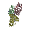
| ||||||||
|---|---|---|---|---|---|---|---|---|---|
| 1 |
| ||||||||
| Unit cell |
|
-Components
-Protein , 2 types, 4 molecules ABCD
| #1: Protein | Mass: 25401.492 Da / Num. of mol.: 2 Source method: isolated from a genetically manipulated source Source: (gene. exp.) Streptomyces sahachiroi (bacteria) / Plasmid: pET Duet-1 / Production host: #2: Protein | Mass: 38937.473 Da / Num. of mol.: 2 Source method: isolated from a genetically manipulated source Source: (gene. exp.) Streptomyces sahachiroi (bacteria) / Plasmid: pET Duet-1 / Production host: |
|---|
-Non-polymers , 5 types, 1082 molecules 


| #3: Chemical | ChemComp-PGE /  Mass: 150.173 Da / Num. of mol.: 1 / Source method: obtained synthetically / Formula: C6H14O4 Mass: 150.173 Da / Num. of mol.: 1 / Source method: obtained synthetically / Formula: C6H14O4 | ||||
|---|---|---|---|---|---|
| #4: Chemical | ChemComp-P4G /  Mass: 162.227 Da / Num. of mol.: 1 / Source method: obtained synthetically / Formula: C8H18O3 Mass: 162.227 Da / Num. of mol.: 1 / Source method: obtained synthetically / Formula: C8H18O3 | ||||
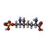
| #5: Chemical |  Mass: 272.276 Da / Num. of mol.: 2 / Source method: obtained synthetically / Formula: C7H16N2O7S / Feature type: SUBJECT OF INVESTIGATION Mass: 272.276 Da / Num. of mol.: 2 / Source method: obtained synthetically / Formula: C7H16N2O7S / Feature type: SUBJECT OF INVESTIGATION#6: Chemical |  Mass: 24.305 Da / Num. of mol.: 3 / Source method: obtained synthetically / Formula: Mg Mass: 24.305 Da / Num. of mol.: 3 / Source method: obtained synthetically / Formula: Mg#7: Water | ChemComp-HOH / | Mass: 18.015 Da / Num. of mol.: 1075 / Source method: isolated from a natural source / Formula: H2O |
-Details
| Has ligand of interest | Y |
|---|
-Experimental details
-Experiment
| Experiment | Method: X-RAY DIFFRACTION / Number of used crystals: 1 |
|---|
- Sample preparation
| Crystal | Density Matthews: 2.22 Å3/Da / Density % sol: 44.47 % |
|---|---|
| Crystal grow | Temperature: 293.15 K / Method: vapor diffusion, hanging drop / pH: 7 / Details: 20% PEG 3350, 0.2M Magnesium formic acid |
-Data collection
| Diffraction | Mean temperature: 95 K / Serial crystal experiment: N |
|---|---|
| Diffraction source | Source: SYNCHROTRON / Site: Photon Factory / Beamline: BL-5A / Wavelength: 1 Å |
| Detector | Type: DECTRIS PILATUS3 6M / Detector: PIXEL / Date: Oct 30, 2021 |
| Radiation | Protocol: SINGLE WAVELENGTH / Monochromatic (M) / Laue (L): M / Scattering type: x-ray |
| Radiation wavelength | Wavelength: 1 Å / Relative weight: 1 |
| Reflection | Resolution: 1.8→48.873 Å / Num. obs: 106322 / % possible obs: 99.6 % / Redundancy: 6.3 % / CC1/2: 0.999 / Rmerge(I) obs: 0.05 / Rpim(I) all: 0.032 / Rrim(I) all: 0.06 / Net I/σ(I): 20.4 |
| Reflection shell | Resolution: 1.8→1.83 Å / Redundancy: 6.5 % / Rmerge(I) obs: 0.26 / Num. unique obs: 5231 / CC1/2: 0.965 / Rpim(I) all: 0.11 / Rrim(I) all: 0.283 |
- Processing
| Software |
| ||||||||||||||||||||||||||||||||||||||||||||||||||||||||||||||||||||||||||||||||||||||||||||||||||||||||||||||||||||||||||||||||||||||||||||||||||||||||||||||||
|---|---|---|---|---|---|---|---|---|---|---|---|---|---|---|---|---|---|---|---|---|---|---|---|---|---|---|---|---|---|---|---|---|---|---|---|---|---|---|---|---|---|---|---|---|---|---|---|---|---|---|---|---|---|---|---|---|---|---|---|---|---|---|---|---|---|---|---|---|---|---|---|---|---|---|---|---|---|---|---|---|---|---|---|---|---|---|---|---|---|---|---|---|---|---|---|---|---|---|---|---|---|---|---|---|---|---|---|---|---|---|---|---|---|---|---|---|---|---|---|---|---|---|---|---|---|---|---|---|---|---|---|---|---|---|---|---|---|---|---|---|---|---|---|---|---|---|---|---|---|---|---|---|---|---|---|---|---|---|---|---|---|
| Refinement | Method to determine structure: SAD / Resolution: 1.8→48.873 Å / Cor.coef. Fo:Fc: 0.962 / Cor.coef. Fo:Fc free: 0.951 / SU B: 2.121 / SU ML: 0.067 / Cross valid method: FREE R-VALUE / ESU R: 0.115 / ESU R Free: 0.108 Details: Hydrogens have been added in their riding positions
| ||||||||||||||||||||||||||||||||||||||||||||||||||||||||||||||||||||||||||||||||||||||||||||||||||||||||||||||||||||||||||||||||||||||||||||||||||||||||||||||||
| Solvent computation | Ion probe radii: 0.8 Å / Shrinkage radii: 0.8 Å / VDW probe radii: 1.2 Å / Solvent model: MASK BULK SOLVENT | ||||||||||||||||||||||||||||||||||||||||||||||||||||||||||||||||||||||||||||||||||||||||||||||||||||||||||||||||||||||||||||||||||||||||||||||||||||||||||||||||
| Displacement parameters | Biso mean: 18.828 Å2
| ||||||||||||||||||||||||||||||||||||||||||||||||||||||||||||||||||||||||||||||||||||||||||||||||||||||||||||||||||||||||||||||||||||||||||||||||||||||||||||||||
| Refinement step | Cycle: LAST / Resolution: 1.8→48.873 Å
| ||||||||||||||||||||||||||||||||||||||||||||||||||||||||||||||||||||||||||||||||||||||||||||||||||||||||||||||||||||||||||||||||||||||||||||||||||||||||||||||||
| Refine LS restraints |
| ||||||||||||||||||||||||||||||||||||||||||||||||||||||||||||||||||||||||||||||||||||||||||||||||||||||||||||||||||||||||||||||||||||||||||||||||||||||||||||||||
| LS refinement shell |
|
