 Movie
Movie Controller

[English] 日本語
 Yorodumi
- PDB-7wst: Cryo-EM structure of the barley Yellow stripe 1 transporter in co... -
+ Open data

- Basic information
| Entry | Database: PDB / ID: 7wst | ||||||
|---|---|---|---|---|---|---|---|
| Title | Cryo-EM structure of the barley Yellow stripe 1 transporter in complex with Fe(III)-DMA | ||||||
 Components Components | Iron-phytosiderophore transporter | ||||||
 Keywords Keywords | METAL TRANSPORT / iron-phytosiderophore transporter / deoxymugineic acid | ||||||
| Function / homology |  Function and homology information Function and homology informationiron-nicotianamine transmembrane transporter activity / oligopeptide transmembrane transporter activity / seed development / response to iron ion / plasma membrane Similarity search - Function | ||||||
| Biological species |  | ||||||
| Method | ELECTRON MICROSCOPY / single particle reconstruction / cryo EM / Resolution: 2.7 Å | ||||||
 Authors Authors | Yamagata, A. | ||||||
| Funding support |  Japan, 1items Japan, 1items
| ||||||
 Citation Citation | Journal: Nat Commun / Year: 2022 Title: Uptake mechanism of iron-phytosiderophore from the soil based on the structure of yellow stripe transporter. Authors: Atsushi Yamagata / Yoshiko Murata / Kosuke Namba / Tohru Terada / Shuya Fukai / Mikako Shirouzu / Abstract: Calcareous soils cover one-third of all land and cause severe growth defects in plants due to the poor water solubility of iron at high pH. Poaceae species use a unique chelation strategy, whereby ...Calcareous soils cover one-third of all land and cause severe growth defects in plants due to the poor water solubility of iron at high pH. Poaceae species use a unique chelation strategy, whereby plants secrete a high-affinity metal chelator, known as phytosiderophores (mugineic acids), and reabsorb the iron-phytosiderophore complex by the yellow stripe 1/yellow stripe 1-like (YS1/YSL) transporter for efficient uptake of iron from the soil. Here, we present three cryo-electron microscopy structures of barley YS1 (HvYS1) in the apo state, in complex with an iron-phytosiderophore complex, Fe(III)-deoxymugineic acid (Fe(III)-DMA), and in complex with the iron-bound synthetic DMA analog (Fe(III)-PDMA). The structures reveal a homodimeric assembly mediated through an anti-parallel β-sheet interaction with cholesterol hemisuccinate. Each protomer adopts an outward open conformation, and Fe(III)-DMA is bound near the extracellular space in the central cavity. Fe(III)-PDMA occupies the same binding site as Fe(III)-DMA, demonstrating that PDMA can function as a potent fertilizer in an essentially identical manner to DMA. Our results provide a structural framework for iron-phytosiderophore recognition and transport by YS1/YSL transporters, which will enable the rational design of new, high-potency fertilizers. | ||||||
| History |
|
- Structure visualization
| Structure viewer | Molecule: MolmilJmol/JSmol |
|---|
- Downloads & links
-Download
| PDBx/mmCIF format | 7wst.cif.gz | 222.5 KB | Display | PDBx/mmCIF format |
|---|---|---|---|---|
| PDB format | pdb7wst.ent.gz | 172.1 KB | Display | PDB format |
| PDBx/mmJSON format | 7wst.json.gz | Tree view | PDBx/mmJSON format | |
| Others |  Other downloads Other downloads |
-Validation report
| Arichive directory | https://data.pdbj.org/pub/pdb/validation_reports/ws/7wstftp://data.pdbj.org/pub/pdb/validation_reports/ws/7wst | HTTPS FTP |
|---|
-Related structure data
| Related structure data |  32766MC 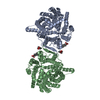 7wsrC 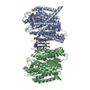 7wsuC M: map data used to model this data C: citing same article ( |
|---|---|
| Similar structure data |


-Links
 PDBj
PDBj- Assembly
| Deposited unit | 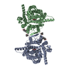
|
|---|---|
| 1 |
|
-Components
| #1: Protein | Mass: 76070.984 Da / Num. of mol.: 2 Source method: isolated from a genetically manipulated source Source: (gene. exp.)   Spodoptera frugiperda (fall armyworm) / References: UniProt: Q2PGC4 Spodoptera frugiperda (fall armyworm) / References: UniProt: Q2PGC4#2: Chemical | ChemComp-Y01 /   Mass: 486.726 Da / Num. of mol.: 4 / Source method: obtained synthetically / Formula: C31H50O4 Mass: 486.726 Da / Num. of mol.: 4 / Source method: obtained synthetically / Formula: C31H50O4#3: Chemical |   Mass: 55.845 Da / Num. of mol.: 2 / Source method: obtained synthetically / Formula: Fe Mass: 55.845 Da / Num. of mol.: 2 / Source method: obtained synthetically / Formula: Fe#4: Chemical |   Mass: 304.296 Da / Num. of mol.: 2 / Source method: obtained synthetically / Formula: C12H20N2O7 Mass: 304.296 Da / Num. of mol.: 2 / Source method: obtained synthetically / Formula: C12H20N2O7Has ligand of interest | N | Has protein modification | Y | |
|---|
-Experimental details
-Experiment
| Experiment | Method: ELECTRON MICROSCOPY |
|---|---|
| EM experiment | Aggregation state: PARTICLE / 3D reconstruction method: single particle reconstruction |
- Sample preparation
| Component | Name: Yellow stripe 1 in the apo state / Type: CELL / Entity ID: #1 / Source: RECOMBINANT |
|---|---|
| Source (natural) | Organism: |
| Source (recombinant) | Organism: Spodoptera frugiperda (fall armyworm) |
| Buffer solution | pH: 7.5 |
| Specimen | Embedding applied: NO / Shadowing applied: NO / Staining applied: NO / Vitrification applied: YES |
| Vitrification | Instrument: FEI VITROBOT MARK IV / Cryogen name: ETHANE / Humidity: 100 % / Chamber temperature: 277 K |
- Electron microscopy imaging
| Experimental equipment |  Model: Titan Krios / Image courtesy: FEI Company |
|---|---|
| Microscopy | Model: FEI TITAN KRIOS |
| Electron gun | Electron source:  FIELD EMISSION GUN / Accelerating voltage: 300 kV / Illumination mode: FLOOD BEAM FIELD EMISSION GUN / Accelerating voltage: 300 kV / Illumination mode: FLOOD BEAM |
| Electron lens | Mode: BRIGHT FIELD / Nominal defocus max: 2500 nm / Nominal defocus min: 600 nm |
| Image recording | Electron dose: 50 e/Å2 / Film or detector model: GATAN K3 BIOQUANTUM (6k x 4k) |
- Processing
| Software |
| ||||||||||||||||||||||||
|---|---|---|---|---|---|---|---|---|---|---|---|---|---|---|---|---|---|---|---|---|---|---|---|---|---|
| EM software |
| ||||||||||||||||||||||||
| CTF correction | Type: PHASE FLIPPING AND AMPLITUDE CORRECTION | ||||||||||||||||||||||||
| Symmetry | Point symmetry: C2 (2 fold cyclic) | ||||||||||||||||||||||||
| 3D reconstruction | Resolution: 2.7 Å / Resolution method: FSC 0.143 CUT-OFF / Num. of particles: 471603 / Symmetry type: POINT | ||||||||||||||||||||||||
| Refinement | Cross valid method: NONE Stereochemistry target values: GeoStd + Monomer Library + CDL v1.2 | ||||||||||||||||||||||||
| Displacement parameters | Biso mean: 45.19 Å2 | ||||||||||||||||||||||||
| Refine LS restraints |
|
