 Movie
Movie Controller


+ Open data

- Basic information
| Entry | Database: PDB / ID: 7wrn | ||||||
|---|---|---|---|---|---|---|---|
| Title | ESRP1 RNaseH-qRRM1 tandem domain | ||||||
 Components Components | Epithelial splicing regulatory protein 1 | ||||||
 Keywords Keywords | SPLICING / qRRM domain / RNA binding / circRNA / RNA BINDING PROTEIN / RNase-H | ||||||
| Function / homology |  Function and homology information Function and homology informationregulation of inner ear auditory receptor cell fate specification / FGFR2 alternative splicing / regulation of RNA splicing / RNA splicing / mRNA processing / Signaling by BRAF and RAF1 fusions / nuclear body / ribonucleoprotein complex / mRNA binding / nucleoplasm / nucleus Similarity search - Function | ||||||
| Biological species |  Homo sapiens (human) Homo sapiens (human) | ||||||
| Method |  X-RAY DIFFRACTION / SYNCHROTRON / MOLECULAR REPLACEMENT / Resolution: 1.85 Å X-RAY DIFFRACTION / SYNCHROTRON / MOLECULAR REPLACEMENT / Resolution: 1.85 Å | ||||||
 Authors Authors | Wu, B.X. / Guo, W.T. / Patel, D.J. | ||||||
| Funding support | 1items
| ||||||
 Citation Citation | Journal: To Be Published Title: ESRP1 RNaseH-qRRM1 tandem domain Authors: Wu, B.X. / Guo, W.T. / Patel, D.J. | ||||||
| History |
|
- Structure visualization
| Structure viewer | Molecule: MolmilJmol/JSmol |
|---|
- Downloads & links
-Download
| PDBx/mmCIF format | 7wrn.cif.gz | 162.8 KB | Display | PDBx/mmCIF format |
|---|---|---|---|---|
| PDB format | pdb7wrn.ent.gz | 106.3 KB | Display | PDB format |
| PDBx/mmJSON format | 7wrn.json.gz | Tree view | PDBx/mmJSON format | |
| Others |  Other downloads Other downloads |
-Validation report
| Arichive directory | https://data.pdbj.org/pub/pdb/validation_reports/wr/7wrnftp://data.pdbj.org/pub/pdb/validation_reports/wr/7wrn | HTTPS FTP |
|---|
-Related structure data
| Related structure data | 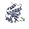 7vkiS S: Starting model for refinement |
|---|---|
| Similar structure data |
-Links
 PDBj
PDBj- Assembly
| Deposited unit | 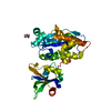
| ||||||||||||
|---|---|---|---|---|---|---|---|---|---|---|---|---|---|
| 1 |
| ||||||||||||
| Unit cell |
|
-Components
| #1: Protein | Mass: 34767.449 Da / Num. of mol.: 1 Source method: isolated from a genetically manipulated source Source: (gene. exp.) Homo sapiens (human) / Gene: ESRP1, RBM35A / Production host:  |
|---|---|
| #2: Chemical | ChemComp-GOL /   Mass: 92.094 Da / Num. of mol.: 1 / Source method: obtained synthetically / Formula: C3H8O3 Mass: 92.094 Da / Num. of mol.: 1 / Source method: obtained synthetically / Formula: C3H8O3 |
| #3: Water | ChemComp-HOH /  Mass: 18.015 Da / Num. of mol.: 125 / Source method: isolated from a natural source / Formula: H2O Mass: 18.015 Da / Num. of mol.: 125 / Source method: isolated from a natural source / Formula: H2O |
| Has ligand of interest | N |
-Experimental details
-Experiment
| Experiment | Method: X-RAY DIFFRACTION / Number of used crystals: 1 |
|---|
- Sample preparation
| Crystal | Density Matthews: 2.03 Å3/Da / Density % sol: 39.39 % |
|---|---|
| Crystal grow | Temperature: 293 K / Method: vapor diffusion, hanging drop Details: 0.1 M Potassium thiocyanate, 30% w/v Polyethylene glycol monomethyl ether 2000 |
-Data collection
| Diffraction | Mean temperature: 100 K / Serial crystal experiment: N |
|---|---|
| Diffraction source | Source: SYNCHROTRON / Site: SSRF  / Beamline: BL19U1 / Wavelength: 0.97852 Å / Beamline: BL19U1 / Wavelength: 0.97852 Å |
| Detector | Type: DECTRIS PILATUS 6M / Detector: PIXEL / Date: Jan 21, 2022 |
| Radiation | Protocol: SINGLE WAVELENGTH / Monochromatic (M) / Laue (L): M / Scattering type: x-ray |
| Radiation wavelength | Wavelength: 0.97852 Å / Relative weight: 1 |
| Reflection | Resolution: 1.85→30 Å / Num. obs: 25216 / % possible obs: 100 % / Redundancy: 25.4 % / Biso Wilson estimate: 24.49 Å2 / CC1/2: 1 / Rmerge(I) obs: 0.108 / Rpim(I) all: 0.022 / Rrim(I) all: 0.111 / Net I/σ(I): 26.9 |
| Reflection shell | Resolution: 1.85→1.95 Å / Redundancy: 25.6 % / Rmerge(I) obs: 1.084 / Mean I/σ(I) obs: 3.7 / Num. unique obs: 3581 / CC1/2: 0.894 / Rpim(I) all: 0.217 / Rrim(I) all: 1.106 / % possible all: 100 |
- Processing
| Software |
| ||||||||||||||||||||||||||||||||||||||||||||||||||||||||||||||||||||||
|---|---|---|---|---|---|---|---|---|---|---|---|---|---|---|---|---|---|---|---|---|---|---|---|---|---|---|---|---|---|---|---|---|---|---|---|---|---|---|---|---|---|---|---|---|---|---|---|---|---|---|---|---|---|---|---|---|---|---|---|---|---|---|---|---|---|---|---|---|---|---|---|
| Refinement | Method to determine structure: MOLECULAR REPLACEMENT Starting model: 7VKI Resolution: 1.85→29.94 Å / SU ML: 0.1636 / Cross valid method: FREE R-VALUE / σ(F): 1.34 / Phase error: 22.836 Stereochemistry target values: GeoStd + Monomer Library + CDL v1.2
| ||||||||||||||||||||||||||||||||||||||||||||||||||||||||||||||||||||||
| Solvent computation | Shrinkage radii: 0.9 Å / VDW probe radii: 1.11 Å / Solvent model: FLAT BULK SOLVENT MODEL | ||||||||||||||||||||||||||||||||||||||||||||||||||||||||||||||||||||||
| Displacement parameters | Biso mean: 31.16 Å2 | ||||||||||||||||||||||||||||||||||||||||||||||||||||||||||||||||||||||
| Refinement step | Cycle: LAST / Resolution: 1.85→29.94 Å
| ||||||||||||||||||||||||||||||||||||||||||||||||||||||||||||||||||||||
| Refine LS restraints |
| ||||||||||||||||||||||||||||||||||||||||||||||||||||||||||||||||||||||
| LS refinement shell |
| ||||||||||||||||||||||||||||||||||||||||||||||||||||||||||||||||||||||
| Refinement TLS params. | Method: refined / Origin x: -7.84778518298 Å / Origin y: -20.9606789864 Å / Origin z: -28.2923155271 Å
| ||||||||||||||||||||||||||||||||||||||||||||||||||||||||||||||||||||||
| Refinement TLS group | Selection details: all |
