 Movie
Movie Controller

[English] 日本語
 Yorodumi
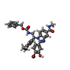
- PDB-7wpn: Methionyl-tRNA synthetase from Staphylococcus aureus complexed wi... -
+ Open data

- Basic information
| Entry | Database: PDB / ID: 7wpn | |||||||||
|---|---|---|---|---|---|---|---|---|---|---|
| Title | Methionyl-tRNA synthetase from Staphylococcus aureus complexed with a phenylbenzimidazole inhibitor and ATP | |||||||||
 Components Components | Methionine--tRNA ligase | |||||||||
 Keywords Keywords | LIGASE/INHIBITOR / LIGASE-INHIBITOR COMPLEX | |||||||||
| Function / homology |  Function and homology information Function and homology informationmethionine-tRNA ligase / methionine-tRNA ligase activity / methionyl-tRNA aminoacylation / tRNA binding / ATP binding / cytoplasm Similarity search - Function | |||||||||
| Biological species |  Staphylococcus aureus subsp. aureus COL (bacteria) Staphylococcus aureus subsp. aureus COL (bacteria) | |||||||||
| Method |  X-RAY DIFFRACTION / MOLECULAR REPLACEMENT / Resolution: 2.4 Å X-RAY DIFFRACTION / MOLECULAR REPLACEMENT / Resolution: 2.4 Å | |||||||||
 Authors Authors | Yi, J. / Cai, Z. / Qiu, H. / Lu, F. / Chen, B. / Luo, Z. / Gu, Q. / Xu, J. / Zhou, H. | |||||||||
| Funding support |  China, 2items China, 2items
| |||||||||
 Citation Citation | Journal: Nucleic Acids Res. / Year: 2022 Title: Fragment screening and structural analyses highlight the ATP-assisted ligand binding for inhibitor discovery against type 1 methionyl-tRNA synthetase. Authors: Yi, J. / Cai, Z. / Qiu, H. / Lu, F. / Luo, Z. / Chen, B. / Gu, Q. / Xu, J. / Zhou, H. | |||||||||
| History |
|
- Structure visualization
| Structure viewer | Molecule: MolmilJmol/JSmol |
|---|
- Downloads & links
-Download
| PDBx/mmCIF format | 7wpn.cif.gz | 114.6 KB | Display | PDBx/mmCIF format |
|---|---|---|---|---|
| PDB format | pdb7wpn.ent.gz | 83.7 KB | Display | PDB format |
| PDBx/mmJSON format | 7wpn.json.gz | Tree view | PDBx/mmJSON format | |
| Others |  Other downloads Other downloads |
-Validation report
| Summary document | 7wpn_validation.pdf.gz | 1.1 MB | Display | wwPDB validaton report |
|---|---|---|---|---|
| Full document | 7wpn_full_validation.pdf.gz | 1.1 MB | Display | |
| Data in XML | 7wpn_validation.xml.gz | 18.3 KB | Display | |
| Data in CIF | 7wpn_validation.cif.gz | 25.1 KB | Display | |
| Arichive directory | https://data.pdbj.org/pub/pdb/validation_reports/wp/7wpnftp://data.pdbj.org/pub/pdb/validation_reports/wp/7wpn | HTTPS FTP |
-Related structure data
| Related structure data | 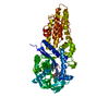 7wpiC 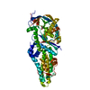 7wpjC 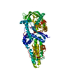 7wpkC 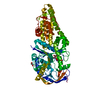 7wplC 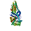 7wpmSC 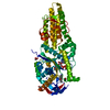 7wptC  7wpxC 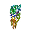 7wq0C S: Starting model for refinement C: citing same article ( |
|---|---|
| Similar structure data |
-Links
 PDBj
PDBj


- Assembly
| Deposited unit | 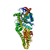
| ||||||||
|---|---|---|---|---|---|---|---|---|---|
| 1 |
| ||||||||
| Unit cell |
|
-Components
-Protein , 1 types, 1 molecules A
| #1: Protein | Mass: 61103.969 Da / Num. of mol.: 1 Source method: isolated from a genetically manipulated source Source: (gene. exp.) Staphylococcus aureus subsp. aureus COL (bacteria)Strain: COL / Gene: metG / Production host: |
|---|
-Non-polymers , 5 types, 32 molecules 


| #2: Chemical | ChemComp-ATP /  Mass: 507.181 Da / Num. of mol.: 1 / Source method: obtained synthetically / Formula: C10H16N5O13P3 / Feature type: SUBJECT OF INVESTIGATION / Comment: ATP, energy-carrying molecule*YM Mass: 507.181 Da / Num. of mol.: 1 / Source method: obtained synthetically / Formula: C10H16N5O13P3 / Feature type: SUBJECT OF INVESTIGATION / Comment: ATP, energy-carrying molecule*YM |
|---|---|
| #3: Chemical | ChemComp-EDO /  Mass: 62.068 Da / Num. of mol.: 1 / Source method: obtained synthetically / Formula: C2H6O2 Mass: 62.068 Da / Num. of mol.: 1 / Source method: obtained synthetically / Formula: C2H6O2 |
| #4: Chemical | ChemComp-29I / ( Mass: 659.526 Da / Num. of mol.: 1 / Source method: obtained synthetically / Formula: C33H31BrN4O6 / Feature type: SUBJECT OF INVESTIGATION Mass: 659.526 Da / Num. of mol.: 1 / Source method: obtained synthetically / Formula: C33H31BrN4O6 / Feature type: SUBJECT OF INVESTIGATION |
| #5: Chemical | ChemComp-ACY /  Mass: 60.052 Da / Num. of mol.: 1 / Source method: obtained synthetically / Formula: C2H4O2 Mass: 60.052 Da / Num. of mol.: 1 / Source method: obtained synthetically / Formula: C2H4O2 |
| #6: Water | ChemComp-HOH / Mass: 18.015 Da / Num. of mol.: 28 / Source method: isolated from a natural source / Formula: H2O |
-Details
| Has ligand of interest | Y |
|---|
-Experimental details
-Experiment
| Experiment | Method: X-RAY DIFFRACTION / Number of used crystals: 1 |
|---|
- Sample preparation
| Crystal | Density Matthews: 2.06 Å3/Da / Density % sol: 40.27 % |
|---|---|
| Crystal grow | Temperature: 300 K / Method: vapor diffusion, sitting drop / pH: 6.5 / Details: Sodium cacodylate, PEG 3350 |
-Data collection
| Diffraction | Mean temperature: 100 K / Serial crystal experiment: N |
|---|---|
| Diffraction source | Source: ROTATING ANODE / Type: RIGAKU MICROMAX-007 HF / Wavelength: 1.5418 Å |
| Detector | Type: RIGAKU HyPix-6000HE / Detector: PIXEL / Date: May 21, 2021 |
| Radiation | Protocol: SINGLE WAVELENGTH / Monochromatic (M) / Laue (L): M / Scattering type: x-ray |
| Radiation wavelength | Wavelength: 1.5418 Å / Relative weight: 1 |
| Reflection | Resolution: 2.4→61.13 Å / Num. obs: 20367 / % possible obs: 99.9 % / Redundancy: 4.7 % / Rmerge(I) obs: 0.14 / Net I/σ(I): 10.6 |
| Reflection shell | Resolution: 2.4→2.49 Å / Rmerge(I) obs: 0.54 / Mean I/σ(I) obs: 3.1 / Num. unique obs: 2082 / % possible all: 99.8 |
- Processing
| Software |
| ||||||||||||||||||||||||||||||||||||||||||||||||||||||||||||
|---|---|---|---|---|---|---|---|---|---|---|---|---|---|---|---|---|---|---|---|---|---|---|---|---|---|---|---|---|---|---|---|---|---|---|---|---|---|---|---|---|---|---|---|---|---|---|---|---|---|---|---|---|---|---|---|---|---|---|---|---|---|
| Refinement | Method to determine structure: MOLECULAR REPLACEMENT Starting model: 7WPM Resolution: 2.4→61.13 Å / Cor.coef. Fo:Fc: 0.917 / Cor.coef. Fo:Fc free: 0.879 / SU B: 9.511 / SU ML: 0.218 / Cross valid method: THROUGHOUT / σ(F): 0 / ESU R: 0.574 / ESU R Free: 0.288 / Stereochemistry target values: MAXIMUM LIKELIHOOD Details: HYDROGENS HAVE BEEN ADDED IN THE RIDING POSITIONS U VALUES : REFINED INDIVIDUALLY
| ||||||||||||||||||||||||||||||||||||||||||||||||||||||||||||
| Solvent computation | Ion probe radii: 0.8 Å / Shrinkage radii: 0.8 Å / VDW probe radii: 1.2 Å / Solvent model: MASK | ||||||||||||||||||||||||||||||||||||||||||||||||||||||||||||
| Displacement parameters | Biso max: 63.24 Å2 / Biso mean: 21.865 Å2 / Biso min: 7.94 Å2
| ||||||||||||||||||||||||||||||||||||||||||||||||||||||||||||
| Refinement step | Cycle: final / Resolution: 2.4→61.13 Å
| ||||||||||||||||||||||||||||||||||||||||||||||||||||||||||||
| Refine LS restraints |
| ||||||||||||||||||||||||||||||||||||||||||||||||||||||||||||
| LS refinement shell | Resolution: 2.4→2.462 Å / Rfactor Rfree error: 0 / Total num. of bins used: 20
|
