 Movie
Movie Controller

[English] 日本語
 Yorodumi
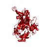
- PDB-7wno: Crystallographic structure of copper amine oxidase from Arthrobac... -
+ Open data

- Basic information
| Entry | Database: PDB / ID: 7wno | ||||||
|---|---|---|---|---|---|---|---|
| Title | Crystallographic structure of copper amine oxidase from Arthrobacter glibiformis at pD 7.4 determined by only neutron diffraction data. | ||||||
 Components Components | Phenylethylamine oxidase | ||||||
 Keywords Keywords | OXIDOREDUCTASE / topaquinone / TPQ | ||||||
| Function / homology |  Function and homology information Function and homology informationprimary-amine oxidase / primary methylamine oxidase activity / amine metabolic process / quinone binding / copper ion binding Similarity search - Function | ||||||
| Biological species |  Arthrobacter globiformis (bacteria) Arthrobacter globiformis (bacteria) | ||||||
| Method | NEUTRON DIFFRACTION /  MIR / Resolution: 1.72 Å MIR / Resolution: 1.72 Å | ||||||
 Authors Authors | Murakawa, T. / Okajima, T. | ||||||
| Funding support |  Japan, 1items Japan, 1items
| ||||||
 Citation Citation | Journal: Iucrj / Year: 2022 Title: Re-evaluation of protein neutron crystallography with and without X-ray/neutron joint refinement. Authors: Murakawa, T. / Kurihara, K. / Adachi, M. / Kusaka, K. / Tanizawa, K. / Okajima, T. | ||||||
| History |
|
- Structure visualization
| Structure viewer | Molecule: MolmilJmol/JSmol |
|---|
- Downloads & links
-Download
| PDBx/mmCIF format | 7wno.cif.gz | 327.5 KB | Display | PDBx/mmCIF format |
|---|---|---|---|---|
| PDB format | pdb7wno.ent.gz | 279.9 KB | Display | PDB format |
| PDBx/mmJSON format | 7wno.json.gz | Tree view | PDBx/mmJSON format | |
| Others |  Other downloads Other downloads |
-Validation report
| Summary document | 7wno_validation.pdf.gz | 336.3 KB | Display | wwPDB validaton report |
|---|---|---|---|---|
| Full document | 7wno_full_validation.pdf.gz | 338.5 KB | Display | |
| Data in XML | 7wno_validation.xml.gz | 14.9 KB | Display | |
| Data in CIF | 7wno_validation.cif.gz | 30.7 KB | Display | |
| Arichive directory | https://data.pdbj.org/pub/pdb/validation_reports/wn/7wnoftp://data.pdbj.org/pub/pdb/validation_reports/wn/7wno | HTTPS FTP |
-Related structure data
-Links
 PDBj
PDBj

- Assembly
| Deposited unit | 
| ||||||||||||||||||
|---|---|---|---|---|---|---|---|---|---|---|---|---|---|---|---|---|---|---|---|
| 1 | 
| ||||||||||||||||||
| Unit cell |
| ||||||||||||||||||
| Components on special symmetry positions |
|
-Components
| #1: Protein | Mass: 68993.836 Da / Num. of mol.: 1 Source method: isolated from a genetically manipulated source Source: (gene. exp.) Arthrobacter globiformis (bacteria) / Production host: |
|---|---|
| #2: Chemical | ChemComp-CU /   Mass: 63.546 Da / Num. of mol.: 1 / Source method: obtained synthetically / Formula: Cu Mass: 63.546 Da / Num. of mol.: 1 / Source method: obtained synthetically / Formula: Cu |
| #3: Chemical | ChemComp-DOD /   Mass: 18.015 Da / Num. of mol.: 1114 / Source method: isolated from a natural source / Formula: D2O Mass: 18.015 Da / Num. of mol.: 1114 / Source method: isolated from a natural source / Formula: D2O |
| Has ligand of interest | Y |
-Experimental details
-Experiment
| Experiment | Method: NEUTRON DIFFRACTION / Number of used crystals: 1 |
|---|
- Sample preparation
| Crystal grow | Temperature: 289 K / Method: microdialysis Details: 1.05-M potassium sodium (Na) tartrate in 25-mM 4-(2-hydroxyethyl)-1-piperazineethanesulfonic acid buffer |
|---|
-Data collection
| Diffraction | Mean temperature: 100 K / Serial crystal experiment: N | |||||||||
|---|---|---|---|---|---|---|---|---|---|---|
| Diffraction source | Source: SPALLATION SOURCE / Site: JPARC MLF / Beamline: BL-03 / Type: J-PARC MLF BEAMLINE BL-03 / Wavelength: 3.0-5.7 | |||||||||
| Detector | Type: iBIX / Detector: DIFFRACTOMETER / Date: Nov 10, 2015 | |||||||||
| Radiation | Protocol: LAUE / Monochromatic (M) / Laue (L): L / Scattering type: neutron | |||||||||
| Radiation wavelength |
| |||||||||
| Reflection | Resolution: 1.72→20.94 Å / Num. obs: 76306 / % possible obs: 87.4 % / Redundancy: 2.664 % / Net I/σ(I): 4.99 | |||||||||
| Reflection shell | Resolution: 1.72→1.78 Å |
- Processing
| Software |
| ||||||||||||||||||||||||||||||||||||||||||||||||||||||||||||||||||||||||||||||||||||||||||||||||||||||||||||||||||||||||||||||||||||||||||||||||||||||||||||||||||||||||||||||||||||||||||||||||||||
|---|---|---|---|---|---|---|---|---|---|---|---|---|---|---|---|---|---|---|---|---|---|---|---|---|---|---|---|---|---|---|---|---|---|---|---|---|---|---|---|---|---|---|---|---|---|---|---|---|---|---|---|---|---|---|---|---|---|---|---|---|---|---|---|---|---|---|---|---|---|---|---|---|---|---|---|---|---|---|---|---|---|---|---|---|---|---|---|---|---|---|---|---|---|---|---|---|---|---|---|---|---|---|---|---|---|---|---|---|---|---|---|---|---|---|---|---|---|---|---|---|---|---|---|---|---|---|---|---|---|---|---|---|---|---|---|---|---|---|---|---|---|---|---|---|---|---|---|---|---|---|---|---|---|---|---|---|---|---|---|---|---|---|---|---|---|---|---|---|---|---|---|---|---|---|---|---|---|---|---|---|---|---|---|---|---|---|---|---|---|---|---|---|---|---|---|---|---|
| Refinement | Method to determine structure: MIR / Resolution: 1.72→20.94 Å / SU ML: 0.27 / Cross valid method: FREE R-VALUE / σ(F): 1.39 / Phase error: 25.33 / Stereochemistry target values: ML
| ||||||||||||||||||||||||||||||||||||||||||||||||||||||||||||||||||||||||||||||||||||||||||||||||||||||||||||||||||||||||||||||||||||||||||||||||||||||||||||||||||||||||||||||||||||||||||||||||||||
| Solvent computation | Shrinkage radii: 0.9 Å / VDW probe radii: 1.11 Å / Solvent model: FLAT BULK SOLVENT MODEL | ||||||||||||||||||||||||||||||||||||||||||||||||||||||||||||||||||||||||||||||||||||||||||||||||||||||||||||||||||||||||||||||||||||||||||||||||||||||||||||||||||||||||||||||||||||||||||||||||||||
| Refine LS restraints |
| ||||||||||||||||||||||||||||||||||||||||||||||||||||||||||||||||||||||||||||||||||||||||||||||||||||||||||||||||||||||||||||||||||||||||||||||||||||||||||||||||||||||||||||||||||||||||||||||||||||
| LS refinement shell |
|
