 Movie
Movie Controller

[English] 日本語
 Yorodumi
- PDB-7wnl: Crystal structure of a mutant Staphylococcus equorum manganese su... -
+ Open data

- Basic information
| Entry | Database: PDB / ID: 7wnl | ||||||
|---|---|---|---|---|---|---|---|
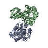
| Title | Crystal structure of a mutant Staphylococcus equorum manganese superoxide dismutase K38R and A121Y | ||||||
 Components Components | Superoxide dismutase | ||||||
 Keywords Keywords | OXIDOREDUCTASE / superoxide dismutase / Staphylococcus equorum | ||||||
| Function / homology |  Function and homology information Function and homology informationsuperoxide dismutase / superoxide dismutase activity / metal ion binding / cytoplasm Similarity search - Function | ||||||
| Biological species |  Staphylococcus equorum (bacteria) Staphylococcus equorum (bacteria) | ||||||
| Method |  X-RAY DIFFRACTION / SYNCHROTRON / MOLECULAR REPLACEMENT / Resolution: 1.51 Å X-RAY DIFFRACTION / SYNCHROTRON / MOLECULAR REPLACEMENT / Resolution: 1.51 Å | ||||||
 Authors Authors | Retnoningrum, D.S. / Yoshida, H. / Artarini, A.A. / Ismaya, W.T. | ||||||
| Funding support | Indonesia, 1items
| ||||||
 Citation Citation | Journal: Appl.Biochem.Biotechnol. / Year: 2023 Title: Introducing Intermolecular Interaction to Strengthen the Stability of MnSOD Dimer. Authors: Retnoningrum, D.S. / Yoshida, H. / Pajatiwi, I. / Muliadi, R. / Utami, R.A. / Artarini, A. / Ismaya, W.T. #1: Journal: Acta Crystallogr F Struct Biol Commun / Year: 2018Title: The first crystal structure of manganese superoxide dismutase from the genus Staphylococcus. Authors: Retnoningrum, D.S. / Yoshida, H. / Arumsari, S. / Kamitori, S. / Ismaya, W.T. | ||||||
| History |
|
- Structure visualization
| Structure viewer | Molecule: MolmilJmol/JSmol |
|---|
- Downloads & links
-Download
| PDBx/mmCIF format | 7wnl.cif.gz | 384.7 KB | Display | PDBx/mmCIF format |
|---|---|---|---|---|
| PDB format | pdb7wnl.ent.gz | 313.5 KB | Display | PDB format |
| PDBx/mmJSON format | 7wnl.json.gz | Tree view | PDBx/mmJSON format | |
| Others |  Other downloads Other downloads |
-Validation report
| Arichive directory | https://data.pdbj.org/pub/pdb/validation_reports/wn/7wnlftp://data.pdbj.org/pub/pdb/validation_reports/wn/7wnl | HTTPS FTP |
|---|
-Related structure data
| Related structure data |  7w6wC  7wnkC  5x2jS S: Starting model for refinement C: citing same article ( |
|---|---|
| Similar structure data |

-Links
 PDBj
PDBj


- Assembly
| Deposited unit | 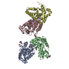
| |||||||||
|---|---|---|---|---|---|---|---|---|---|---|
| 1 | 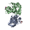
| |||||||||
| 2 | 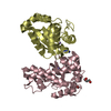
| |||||||||
| Unit cell |
| |||||||||
| Components on special symmetry positions |
|
-Components
-Protein , 1 types, 4 molecules ABCD
| #1: Protein | Mass: 23622.139 Da / Num. of mol.: 4 / Mutation: D13R, K38R, A121Y Source method: isolated from a genetically manipulated source Source: (gene. exp.) Staphylococcus equorum (bacteria) / Gene: AST02_02815 / Plasmid: plasmid / Details (production host): pJExpress414 / Production host: |
|---|
-Non-polymers , 6 types, 1176 molecules 


| #2: Chemical |  Mass: 106.120 Da / Num. of mol.: 2 / Source method: obtained synthetically / Formula: C4H10O3 Mass: 106.120 Da / Num. of mol.: 2 / Source method: obtained synthetically / Formula: C4H10O3#3: Chemical | ChemComp-MN /  Mass: 54.938 Da / Num. of mol.: 4 / Source method: obtained synthetically / Formula: Mn Mass: 54.938 Da / Num. of mol.: 4 / Source method: obtained synthetically / Formula: Mn#4: Chemical |  Mass: 79.904 Da / Num. of mol.: 2 / Source method: obtained synthetically / Formula: Br Mass: 79.904 Da / Num. of mol.: 2 / Source method: obtained synthetically / Formula: Br#5: Chemical | ChemComp-TRS / |  Mass: 122.143 Da / Num. of mol.: 1 / Source method: obtained synthetically / Formula: C4H12NO3 / Comment: pH buffer*YM Mass: 122.143 Da / Num. of mol.: 1 / Source method: obtained synthetically / Formula: C4H12NO3 / Comment: pH buffer*YM#6: Chemical | ChemComp-AZI / |  Mass: 42.020 Da / Num. of mol.: 1 / Source method: obtained synthetically / Formula: N3 Mass: 42.020 Da / Num. of mol.: 1 / Source method: obtained synthetically / Formula: N3#7: Water | ChemComp-HOH / | Mass: 18.015 Da / Num. of mol.: 1166 / Source method: isolated from a natural source / Formula: H2O |
|---|
-Details
| Has ligand of interest | N |
|---|
-Experimental details
-Experiment
| Experiment | Method: X-RAY DIFFRACTION / Number of used crystals: 1 |
|---|
- Sample preparation
| Crystal | Density Matthews: 2.45 Å3/Da / Density % sol: 49.85 % |
|---|---|
| Crystal grow | Temperature: 293 K / Method: vapor diffusion, sitting drop / pH: 6.5 Details: sodium fluoride, sodium bromide, sodium iodide, imidazole, MES, PEG MME 500, PEG 20000 |
-Data collection
| Diffraction | Mean temperature: 100 K / Serial crystal experiment: N | ||||||||||||||||||||||||||||||
|---|---|---|---|---|---|---|---|---|---|---|---|---|---|---|---|---|---|---|---|---|---|---|---|---|---|---|---|---|---|---|---|
| Diffraction source | Source: SYNCHROTRON / Site: Photon Factory  / Beamline: BL-5A / Wavelength: 0.98 Å / Beamline: BL-5A / Wavelength: 0.98 Å | ||||||||||||||||||||||||||||||
| Detector | Type: DECTRIS PILATUS3 S 6M / Detector: PIXEL / Date: Jun 5, 2021 | ||||||||||||||||||||||||||||||
| Radiation | Protocol: SINGLE WAVELENGTH / Monochromatic (M) / Laue (L): M / Scattering type: x-ray | ||||||||||||||||||||||||||||||
| Radiation wavelength | Wavelength: 0.98 Å / Relative weight: 1 | ||||||||||||||||||||||||||||||
| Reflection | Resolution: 1.51→45.93 Å / Num. obs: 144687 / % possible obs: 99.8 % / Redundancy: 6 % / CC1/2: 0.997 / Rmerge(I) obs: 0.105 / Rpim(I) all: 0.047 / Rrim(I) all: 0.115 / Net I/σ(I): 10.7 / Num. measured all: 873802 / Scaling rejects: 74 | ||||||||||||||||||||||||||||||
| Reflection shell | Diffraction-ID: 1
|
- Processing
| Software |
| |||||||||||||||||||||||||||||||||||||||||||||||||||||||||||||||||
|---|---|---|---|---|---|---|---|---|---|---|---|---|---|---|---|---|---|---|---|---|---|---|---|---|---|---|---|---|---|---|---|---|---|---|---|---|---|---|---|---|---|---|---|---|---|---|---|---|---|---|---|---|---|---|---|---|---|---|---|---|---|---|---|---|---|---|
| Refinement | Method to determine structure: MOLECULAR REPLACEMENT Starting model: 5x2j Resolution: 1.51→44.79 Å / Cor.coef. Fo:Fc: 0.969 / Cor.coef. Fo:Fc free: 0.938 / SU B: 4.911 / SU ML: 0.078 / Cross valid method: THROUGHOUT / σ(F): 0 / ESU R: 0.088 / ESU R Free: 0.088 / Stereochemistry target values: MAXIMUM LIKELIHOOD Details: HYDROGENS HAVE BEEN ADDED IN THE RIDING POSITIONS U VALUES : REFINED INDIVIDUALLY
| |||||||||||||||||||||||||||||||||||||||||||||||||||||||||||||||||
| Solvent computation | Ion probe radii: 0.8 Å / Shrinkage radii: 0.8 Å / VDW probe radii: 1.2 Å / Solvent model: MASK | |||||||||||||||||||||||||||||||||||||||||||||||||||||||||||||||||
| Displacement parameters | Biso max: 105.49 Å2 / Biso mean: 19.993 Å2 / Biso min: 8.08 Å2
| |||||||||||||||||||||||||||||||||||||||||||||||||||||||||||||||||
| Refinement step | Cycle: final / Resolution: 1.51→44.79 Å
| |||||||||||||||||||||||||||||||||||||||||||||||||||||||||||||||||
| Refine LS restraints |
| |||||||||||||||||||||||||||||||||||||||||||||||||||||||||||||||||
| LS refinement shell | Resolution: 1.51→1.549 Å / Rfactor Rfree error: 0 / Total num. of bins used: 20
|
