 Movie
Movie Controller

[English] 日本語
 Yorodumi
- PDB-7wmq: Crystal Structure of the second bromodomain of human BRD2 in comp... -
+ Open data

- Basic information
| Entry | Database: PDB / ID: 7wmq | ||||||
|---|---|---|---|---|---|---|---|
| Title | Crystal Structure of the second bromodomain of human BRD2 in complex with the inhibitor Y13157 | ||||||
 Components Components | Isoform 4 of Bromodomain-containing protein 2 | ||||||
 Keywords Keywords | PROTEIN BINDING / BRD2-BD2 / Bromodomain / Inhibitor | ||||||
| Function / homology |  Function and homology information Function and homology informationacetylation-dependent protein binding / histone H3K14ac reader activity / chromatin looping / histone H4K5ac reader activity / histone H4K12ac reader activity / RUNX3 regulates p14-ARF / positive regulation of T-helper 17 cell lineage commitment / protein localization to chromatin / neural tube closure / nucleosome assembly ...acetylation-dependent protein binding / histone H3K14ac reader activity / chromatin looping / histone H4K5ac reader activity / histone H4K12ac reader activity / RUNX3 regulates p14-ARF / positive regulation of T-helper 17 cell lineage commitment / protein localization to chromatin / neural tube closure / nucleosome assembly / spermatogenesis / histone binding / nuclear speck / chromatin remodeling / protein serine/threonine kinase activity / chromatin binding / regulation of transcription by RNA polymerase II / chromatin / nucleoplasm / nucleus / cytoplasm Similarity search - Function | ||||||
| Biological species |  Homo sapiens (human) Homo sapiens (human) | ||||||
| Method |  X-RAY DIFFRACTION / SYNCHROTRON / MOLECULAR REPLACEMENT / Resolution: 2.37 Å X-RAY DIFFRACTION / SYNCHROTRON / MOLECULAR REPLACEMENT / Resolution: 2.37 Å | ||||||
 Authors Authors | Li, J. / Zhang, C. / Xu, H. / Zhuang, X. / Wu, X. / Zhang, Y. / Xu, Y. | ||||||
| Funding support |  China, 1items China, 1items
| ||||||
 Citation Citation | Journal: J.Med.Chem. / Year: 2022 Title: Structure-Based Discovery and Optimization of Furo[3,2- c ]pyridin-4(5 H )-one Derivatives as Potent and Second Bromodomain (BD2)-Selective Bromo and Extra Terminal Domain (BET) Inhibitors. Authors: Li, J. / Zhang, C. / Xu, H. / Wang, C. / Dong, R. / Shen, H. / Zhuang, X. / Chen, X. / Li, Q. / Lu, J. / Zhang, M. / Wu, X. / Loomes, K.M. / Zhou, Y. / Zhang, Y. / Liu, J. / Xu, Y. | ||||||
| History |
|
- Structure visualization
| Structure viewer | Molecule: MolmilJmol/JSmol |
|---|
- Downloads & links
-Download
| PDBx/mmCIF format | 7wmq.cif.gz | 116 KB | Display | PDBx/mmCIF format |
|---|---|---|---|---|
| PDB format | pdb7wmq.ent.gz | 88.8 KB | Display | PDB format |
| PDBx/mmJSON format | 7wmq.json.gz | Tree view | PDBx/mmJSON format | |
| Others |  Other downloads Other downloads |
-Validation report
| Arichive directory | https://data.pdbj.org/pub/pdb/validation_reports/wm/7wmqftp://data.pdbj.org/pub/pdb/validation_reports/wm/7wmq | HTTPS FTP |
|---|
-Related structure data
| Related structure data | 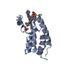 7wjsC 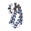 7wkyC 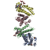 7wlnC 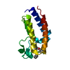 7wmuC 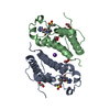 7wn5C 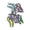 7wnaC 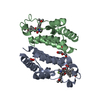 7wniC 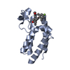 6e6jS S: Starting model for refinement C: citing same article ( |
|---|---|
| Similar structure data |
-Links
 PDBj
PDBj
- Assembly
| Deposited unit | 
| ||||||||
|---|---|---|---|---|---|---|---|---|---|
| 1 |
| ||||||||
| Unit cell |
|
-Components
| #1: Protein | Mass: 15981.218 Da / Num. of mol.: 4 Source method: isolated from a genetically manipulated source Source: (gene. exp.) Homo sapiens (human) / Gene: BRD2, KIAA9001, RING3 / Production host:  #2: Chemical | ChemComp-JFL /   Mass: 541.613 Da / Num. of mol.: 4 / Source method: obtained synthetically / Formula: C32H32FN3O4 / Feature type: SUBJECT OF INVESTIGATION Mass: 541.613 Da / Num. of mol.: 4 / Source method: obtained synthetically / Formula: C32H32FN3O4 / Feature type: SUBJECT OF INVESTIGATION#3: Chemical | ChemComp-GOL /   Mass: 92.094 Da / Num. of mol.: 6 / Source method: obtained synthetically / Formula: C3H8O3 Mass: 92.094 Da / Num. of mol.: 6 / Source method: obtained synthetically / Formula: C3H8O3#4: Chemical | ChemComp-FMT /   Mass: 46.025 Da / Num. of mol.: 15 / Source method: obtained synthetically / Formula: CH2O2 Mass: 46.025 Da / Num. of mol.: 15 / Source method: obtained synthetically / Formula: CH2O2#5: Water | ChemComp-HOH / |  Mass: 18.015 Da / Num. of mol.: 210 / Source method: isolated from a natural source / Formula: H2O Mass: 18.015 Da / Num. of mol.: 210 / Source method: isolated from a natural source / Formula: H2OHas ligand of interest | Y | |
|---|
-Experimental details
-Experiment
| Experiment | Method: X-RAY DIFFRACTION / Number of used crystals: 1 |
|---|
- Sample preparation
| Crystal | Density Matthews: 3.7 Å3/Da / Density % sol: 66.74 % |
|---|---|
| Crystal grow | Temperature: 277 K / Method: vapor diffusion, sitting drop Details: 3.5 M Sodium formate, 0.1 M Sodium acetate trihydrate pH 4.6 |
-Data collection
| Diffraction | Mean temperature: 197 K / Serial crystal experiment: N | |||||||||||||||||||||||||||
|---|---|---|---|---|---|---|---|---|---|---|---|---|---|---|---|---|---|---|---|---|---|---|---|---|---|---|---|---|
| Diffraction source | Source: SYNCHROTRON / Site: SSRF / Beamline: BL18U1 / Wavelength: 0.97915 Å | |||||||||||||||||||||||||||
| Detector | Type: DECTRIS PILATUS3 6M / Detector: PIXEL / Date: May 8, 2021 | |||||||||||||||||||||||||||
| Radiation | Protocol: SINGLE WAVELENGTH / Monochromatic (M) / Laue (L): M / Scattering type: x-ray | |||||||||||||||||||||||||||
| Radiation wavelength | Wavelength: 0.97915 Å / Relative weight: 1 | |||||||||||||||||||||||||||
| Reflection | Resolution: 2.37→72.04 Å / Num. obs: 37617 / % possible obs: 95.9 % / Redundancy: 6.1 % / CC1/2: 0.986 / Rmerge(I) obs: 0.189 / Rpim(I) all: 0.083 / Rrim(I) all: 0.207 / Net I/σ(I): 7.2 | |||||||||||||||||||||||||||
| Reflection shell | Diffraction-ID: 1 / Redundancy: 5.8 %
|
- Processing
| Software |
| ||||||||||||||||||||||||||||||||||||||||||||||||||||||||||||
|---|---|---|---|---|---|---|---|---|---|---|---|---|---|---|---|---|---|---|---|---|---|---|---|---|---|---|---|---|---|---|---|---|---|---|---|---|---|---|---|---|---|---|---|---|---|---|---|---|---|---|---|---|---|---|---|---|---|---|---|---|---|
| Refinement | Method to determine structure: MOLECULAR REPLACEMENT Starting model: 6E6J Resolution: 2.37→72.04 Å / Cor.coef. Fo:Fc: 0.944 / Cor.coef. Fo:Fc free: 0.934 / SU B: 5.388 / SU ML: 0.123 / Cross valid method: THROUGHOUT / σ(F): 0 / ESU R: 0.212 / ESU R Free: 0.175 / Stereochemistry target values: MAXIMUM LIKELIHOOD Details: HYDROGENS HAVE BEEN ADDED IN THE RIDING POSITIONS U VALUES : REFINED INDIVIDUALLY
| ||||||||||||||||||||||||||||||||||||||||||||||||||||||||||||
| Solvent computation | Ion probe radii: 0.8 Å / Shrinkage radii: 0.8 Å / VDW probe radii: 1.2 Å / Solvent model: MASK | ||||||||||||||||||||||||||||||||||||||||||||||||||||||||||||
| Displacement parameters | Biso max: 92.84 Å2 / Biso mean: 29.293 Å2 / Biso min: 12.69 Å2
| ||||||||||||||||||||||||||||||||||||||||||||||||||||||||||||
| Refinement step | Cycle: final / Resolution: 2.37→72.04 Å
| ||||||||||||||||||||||||||||||||||||||||||||||||||||||||||||
| Refine LS restraints |
| ||||||||||||||||||||||||||||||||||||||||||||||||||||||||||||
| LS refinement shell | Resolution: 2.37→2.432 Å / Rfactor Rfree error: 0 / Total num. of bins used: 20
|
