[English] 日本語
 Yorodumi
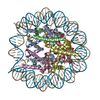
- PDB-7wlr: Cryo-EM structure of the nucleosome containing Komagataella pasto... -
+ Open data
- Basic information
| Entry | Database: PDB / ID: 7wlr | ||||||||||||||||||||||||
|---|---|---|---|---|---|---|---|---|---|---|---|---|---|---|---|---|---|---|---|---|---|---|---|---|---|
| Title | Cryo-EM structure of the nucleosome containing Komagataella pastoris histones | ||||||||||||||||||||||||
 Components Components |
| ||||||||||||||||||||||||
 Keywords Keywords | GENE REGULATION/DNA / Chromatin / Nucleosome / Komagataella pastoris / Histone H2A / Histone H2B / Histone H3 / Histone H4 / DNA / Epigenetic / Gene regulation / cryo-EM / GENE REGULATION-DNA complex | ||||||||||||||||||||||||
| Function / homology |  Function and homology information Function and homology informationstructural constituent of chromatin / nucleosome / protein heterodimerization activity / DNA repair / DNA binding / nucleus Similarity search - Function | ||||||||||||||||||||||||
| Biological species |  Komagataella pastoris (fungus) Komagataella pastoris (fungus)synthetic construct (others) | ||||||||||||||||||||||||
| Method | ELECTRON MICROSCOPY / single particle reconstruction / cryo EM / Resolution: 3.54 Å | ||||||||||||||||||||||||
 Authors Authors | Fukushima, Y. / Hatazawa, S. / Hirai, S. / Kujirai, T. / Takizawa, Y. / Kurumizaka, H. | ||||||||||||||||||||||||
| Funding support |  Japan, 7items Japan, 7items
| ||||||||||||||||||||||||
 Citation Citation |  Journal: J Biochem / Year: 2022 Journal: J Biochem / Year: 2022Title: Structural and biochemical analyses of the nucleosome containing Komagataella pastoris histones. Authors: Yutaro Fukushima / Suguru Hatazawa / Seiya Hirai / Tomoya Kujirai / Haruhiko Ehara / Shun-Ichi Sekine / Yoshimasa Takizawa / Hitoshi Kurumizaka /  Abstract: Komagataella pastoris is a methylotrophic yeast that is commonly used as a host cell for protein production. In the present study, we reconstituted the nucleosome with K. pastoris histones and ...Komagataella pastoris is a methylotrophic yeast that is commonly used as a host cell for protein production. In the present study, we reconstituted the nucleosome with K. pastoris histones and determined the structure of the nucleosome core particle by cryogenic electron microscopy. In the K. pastoris nucleosome, the histones form an octamer and the DNA is left-handedly wrapped around it. Micrococcal nuclease assays revealed that the DNA ends of the K. pastoris nucleosome are somewhat more accessible, as compared with those of the human nucleosome. In vitro transcription assays demonstrated that the K. pastoris nucleosome is transcribed by the K. pastoris RNA polymerase II (RNAPII) more efficiently than the human nucleosome, while the RNAPII pausing positions of the K. pastoris nucleosome are the same as those of the human nucleosome. These results suggested that the DNA end flexibility may enhance the transcription efficiency in the nucleosome but minimally affect the nucleosomal pausing positions of RNAPII. | ||||||||||||||||||||||||
| History |
|
- Structure visualization
| Structure viewer | Molecule:  Molmil Molmil Jmol/JSmol Jmol/JSmol |
|---|
- Downloads & links
- Download
| PDBx/mmCIF format |  7wlr.cif.gz 7wlr.cif.gz | 277.9 KB | Display |  PDBx/mmCIF format PDBx/mmCIF format |
|---|---|---|---|---|
| PDB format |  pdb7wlr.ent.gz pdb7wlr.ent.gz | 209.4 KB | Display |  PDB format PDB format |
| PDBx/mmJSON format |  7wlr.json.gz 7wlr.json.gz | Tree view |  PDBx/mmJSON format PDBx/mmJSON format | |
| Others |  Other downloads Other downloads |
-Validation report
| Arichive directory |  https://data.pdbj.org/pub/pdb/validation_reports/wl/7wlr https://data.pdbj.org/pub/pdb/validation_reports/wl/7wlr ftp://data.pdbj.org/pub/pdb/validation_reports/wl/7wlr ftp://data.pdbj.org/pub/pdb/validation_reports/wl/7wlr | HTTPS FTP |
|---|
-Related structure data
| Related structure data |  32591MC M: map data used to model this data C: citing same article ( |
|---|---|
| Similar structure data | Similarity search - Function & homology  F&H Search F&H Search |
- Links
Links
- Assembly
| Deposited unit | 
|
|---|---|
| 1 |
|
- Components
-Protein , 4 types, 8 molecules AEBFCGDH
| #1: Protein | Mass: 11601.565 Da / Num. of mol.: 2 Source method: isolated from a genetically manipulated source Source: (gene. exp.)  Komagataella pastoris (fungus) / Gene: ATY40_BA7502079, ATY40_BA7504318 / Production host: Komagataella pastoris (fungus) / Gene: ATY40_BA7502079, ATY40_BA7504318 / Production host:  #2: Protein | Mass: 10030.811 Da / Num. of mol.: 2 Source method: isolated from a genetically manipulated source Source: (gene. exp.)  Komagataella pastoris (fungus) / Gene: ATY40_BA7502080, ATY40_BA7504319 / Production host: Komagataella pastoris (fungus) / Gene: ATY40_BA7502080, ATY40_BA7504319 / Production host:  #3: Protein | Mass: 11810.682 Da / Num. of mol.: 2 Source method: isolated from a genetically manipulated source Source: (gene. exp.)  Komagataella pastoris (fungus) / Gene: HTA1, ATY40_BA7502752 / Production host: Komagataella pastoris (fungus) / Gene: HTA1, ATY40_BA7502752 / Production host:  #4: Protein | Mass: 11018.554 Da / Num. of mol.: 2 Source method: isolated from a genetically manipulated source Source: (gene. exp.)  Komagataella pastoris (fungus) / Gene: ATY40_BA7502753 / Production host: Komagataella pastoris (fungus) / Gene: ATY40_BA7502753 / Production host:  |
|---|
-DNA chain , 2 types, 2 molecules IJ
| #5: DNA chain | Mass: 44520.383 Da / Num. of mol.: 1 / Source method: obtained synthetically / Source: (synth.) synthetic construct (others) |
|---|---|
| #6: DNA chain | Mass: 44991.660 Da / Num. of mol.: 1 / Source method: obtained synthetically / Source: (synth.) synthetic construct (others) |
-Experimental details
-Experiment
| Experiment | Method: ELECTRON MICROSCOPY |
|---|---|
| EM experiment | Aggregation state: PARTICLE / 3D reconstruction method: single particle reconstruction |
- Sample preparation
| Component |
| ||||||||||||||||||||||||
|---|---|---|---|---|---|---|---|---|---|---|---|---|---|---|---|---|---|---|---|---|---|---|---|---|---|
| Source (natural) | Organism:  Komagataella pastoris (fungus) Komagataella pastoris (fungus) | ||||||||||||||||||||||||
| Source (recombinant) | Organism:  | ||||||||||||||||||||||||
| Buffer solution | pH: 7.5 | ||||||||||||||||||||||||
| Specimen | Conc.: 1.3 mg/ml / Embedding applied: NO / Shadowing applied: NO / Staining applied: NO / Vitrification applied: YES | ||||||||||||||||||||||||
| Specimen support | Grid material: COPPER / Grid mesh size: 200 divisions/in. / Grid type: Quantifoil R1.2/1.3 | ||||||||||||||||||||||||
| Vitrification | Instrument: FEI VITROBOT MARK IV / Cryogen name: ETHANE / Humidity: 100 % / Chamber temperature: 277 K |
- Electron microscopy imaging
| Experimental equipment |  Model: Titan Krios / Image courtesy: FEI Company |
|---|---|
| Microscopy | Model: FEI TITAN KRIOS |
| Electron gun | Electron source:  FIELD EMISSION GUN / Accelerating voltage: 300 kV / Illumination mode: FLOOD BEAM FIELD EMISSION GUN / Accelerating voltage: 300 kV / Illumination mode: FLOOD BEAM |
| Electron lens | Mode: BRIGHT FIELD / Nominal defocus max: 2500 nm / Nominal defocus min: 1250 nm |
| Image recording | Electron dose: 57 e/Å2 / Film or detector model: GATAN K3 BIOQUANTUM (6k x 4k) |
- Processing
| EM software | Name: RELION / Version: 3.1 / Category: 3D reconstruction |
|---|---|
| CTF correction | Type: PHASE FLIPPING AND AMPLITUDE CORRECTION |
| 3D reconstruction | Resolution: 3.54 Å / Resolution method: FSC 0.143 CUT-OFF / Num. of particles: 181796 / Symmetry type: POINT |
 Movie
Movie Controller


 PDBj
PDBj


