 Movie
Movie Controller


+ Open data

- Basic information
| Entry | Database: PDB / ID: 7wlb | |||||||||||||||
|---|---|---|---|---|---|---|---|---|---|---|---|---|---|---|---|---|
| Title | Mouse Pendrin in chloride and iodide buffer in asymmetric state | |||||||||||||||
 Components Components | Pendrin | |||||||||||||||
 Keywords Keywords | TRANSPORT PROTEIN / exchange / transport / slc | |||||||||||||||
| Function / homology |  Function and homology information Function and homology informationInorganic anion exchange by SLC26 transporters / inorganic anion transport / iodide transmembrane transporter activity / oxalate transmembrane transporter activity / sulfate transmembrane transporter activity / iodide transport / secondary active sulfate transmembrane transporter activity / sulfate transmembrane transport / monoatomic anion transmembrane transporter activity / regulation of pH ...Inorganic anion exchange by SLC26 transporters / inorganic anion transport / iodide transmembrane transporter activity / oxalate transmembrane transporter activity / sulfate transmembrane transporter activity / iodide transport / secondary active sulfate transmembrane transporter activity / sulfate transmembrane transport / monoatomic anion transmembrane transporter activity / regulation of pH / chloride:bicarbonate antiporter activity / chloride transmembrane transporter activity / animal organ morphogenesis / chloride transmembrane transport / brush border membrane / regulation of protein localization / apical plasma membrane / extracellular exosome / membrane / plasma membrane Similarity search - Function | |||||||||||||||
| Biological species |  | |||||||||||||||
| Method | ELECTRON MICROSCOPY / single particle reconstruction / cryo EM / Resolution: 4.1 Å | |||||||||||||||
 Authors Authors | Liu, Q.Y. / Zhang, X. / Sun, L. / Chen, Z.G. | |||||||||||||||
| Funding support |  China, 1items China, 1items
| |||||||||||||||
 Citation Citation | Journal: Nat Commun / Year: 2023 Title: Asymmetric pendrin homodimer reveals its molecular mechanism as anion exchanger. Authors: Qianying Liu / Xiang Zhang / Hui Huang / Yuxin Chen / Fang Wang / Aihua Hao / Wuqiang Zhan / Qiyu Mao / Yuxia Hu / Lin Han / Yifang Sun / Meng Zhang / Zhimin Liu / Geng-Lin Li / Weijia Zhang ...Authors: Qianying Liu / Xiang Zhang / Hui Huang / Yuxin Chen / Fang Wang / Aihua Hao / Wuqiang Zhan / Qiyu Mao / Yuxia Hu / Lin Han / Yifang Sun / Meng Zhang / Zhimin Liu / Geng-Lin Li / Weijia Zhang / Yilai Shu / Lei Sun / Zhenguo Chen / Abstract: Pendrin (SLC26A4) is an anion exchanger expressed in the apical membranes of selected epithelia. Pendrin ablation causes Pendred syndrome, a genetic disorder associated with sensorineural hearing ...Pendrin (SLC26A4) is an anion exchanger expressed in the apical membranes of selected epithelia. Pendrin ablation causes Pendred syndrome, a genetic disorder associated with sensorineural hearing loss, hypothyroid goiter, and reduced blood pressure. However its molecular structure has remained unknown, limiting our understanding of the structural basis of transport. Here, we determine the cryo-electron microscopy structures of mouse pendrin with symmetric and asymmetric homodimer conformations. The asymmetric homodimer consists of one inward-facing protomer and the other outward-facing protomer, representing coincident uptake and secretion- a unique state of pendrin as an electroneutral exchanger. The multiple conformations presented here provide an inverted alternate-access mechanism for anion exchange. The structural and functional data presented here disclose the properties of an anion exchange cleft and help understand the importance of disease-associated variants, which will shed light on the pendrin exchange mechanism. | |||||||||||||||
| History |
|
- Structure visualization
| Structure viewer | Molecule: MolmilJmol/JSmol |
|---|
- Downloads & links
-Download
| PDBx/mmCIF format | 7wlb.cif.gz | 231.8 KB | Display | PDBx/mmCIF format |
|---|---|---|---|---|
| PDB format | pdb7wlb.ent.gz | 185.9 KB | Display | PDB format |
| PDBx/mmJSON format | 7wlb.json.gz | Tree view | PDBx/mmJSON format | |
| Others |  Other downloads Other downloads |
-Validation report
| Arichive directory | https://data.pdbj.org/pub/pdb/validation_reports/wl/7wlbftp://data.pdbj.org/pub/pdb/validation_reports/wl/7wlb | HTTPS FTP |
|---|
-Related structure data
| Related structure data |  32580MC 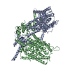 7wk1C  7wk7C 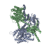 7wl2C  7wl7C 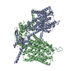 7wl8C 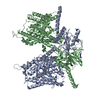 7wl9C  7wlaC 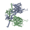 7wleC M: map data used to model this data C: citing same article ( |
|---|---|
| Similar structure data |


-Links
 PDBj
PDBj
- Assembly
| Deposited unit | 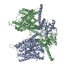
|
|---|---|
| 1 |
|
-Components
| #1: Protein | Mass: 85769.578 Da / Num. of mol.: 2 Source method: isolated from a genetically manipulated source Source: (gene. exp.)  Homo sapiens (human) / References: UniProt: Q9R155 Homo sapiens (human) / References: UniProt: Q9R155Has protein modification | N | |
|---|
-Experimental details
-Experiment
| Experiment | Method: ELECTRON MICROSCOPY |
|---|---|
| EM experiment | Aggregation state: PARTICLE / 3D reconstruction method: single particle reconstruction |
- Sample preparation
| Component | Name: mouse Pendrin in chloride and bicarbonate in inward state Type: COMPLEX / Entity ID: all / Source: RECOMBINANT |
|---|---|
| Source (natural) | Organism: |
| Source (recombinant) | Organism: Homo sapiens (human) |
| Buffer solution | pH: 8 |
| Specimen | Embedding applied: NO / Shadowing applied: NO / Staining applied: NO / Vitrification applied: YES |
| Vitrification | Cryogen name: ETHANE |
- Electron microscopy imaging
| Experimental equipment |  Model: Titan Krios / Image courtesy: FEI Company |
|---|---|
| Microscopy | Model: FEI TITAN KRIOS |
| Electron gun | Electron source:  FIELD EMISSION GUN / Accelerating voltage: 300 kV / Illumination mode: FLOOD BEAM FIELD EMISSION GUN / Accelerating voltage: 300 kV / Illumination mode: FLOOD BEAM |
| Electron lens | Mode: BRIGHT FIELD / Nominal defocus max: 2500 nm / Nominal defocus min: 1200 nm |
| Image recording | Electron dose: 58 e/Å2 / Film or detector model: GATAN K3 (6k x 4k) |
- Processing
| Software | Name: PHENIX / Version: 1.17.1_3660: / Classification: refinement | ||||||||||||||||||||||||
|---|---|---|---|---|---|---|---|---|---|---|---|---|---|---|---|---|---|---|---|---|---|---|---|---|---|
| EM software | Name: PHENIX / Category: model refinement | ||||||||||||||||||||||||
| CTF correction | Type: PHASE FLIPPING AND AMPLITUDE CORRECTION | ||||||||||||||||||||||||
| 3D reconstruction | Resolution: 4.1 Å / Resolution method: FSC 0.143 CUT-OFF / Num. of particles: 45859 / Symmetry type: POINT | ||||||||||||||||||||||||
| Refine LS restraints |
|
