 Movie
Movie Controller


+ Open data

- Basic information
| Entry | Database: PDB / ID: 7wkk | ||||||
|---|---|---|---|---|---|---|---|
| Title | Cryo-EM structure of the IR subunit from X. laevis NPC | ||||||
 Components Components |
| ||||||
 Keywords Keywords | STRUCTURAL PROTEIN / nuclear pore complex / inner ring / Nup205 / Nup93 / Nup188 / Nup155 / NDC1 / Aladin | ||||||
| Function / homology |  Function and homology information Function and homology informationnuclear pore transmembrane ring / protein localization to nuclear inner membrane / nuclear pore inner ring / nuclear pore central transport channel / transcription-dependent tethering of RNA polymerase II gene DNA at nuclear periphery / nuclear pore complex assembly / nuclear pore organization / NLS-bearing protein import into nucleus / structural constituent of nuclear pore / nuclear localization sequence binding ...nuclear pore transmembrane ring / protein localization to nuclear inner membrane / nuclear pore inner ring / nuclear pore central transport channel / transcription-dependent tethering of RNA polymerase II gene DNA at nuclear periphery / nuclear pore complex assembly / nuclear pore organization / NLS-bearing protein import into nucleus / structural constituent of nuclear pore / nuclear localization sequence binding / RNA export from nucleus / nucleocytoplasmic transport / poly(A)+ mRNA export from nucleus / mRNA transport / nuclear pore / nuclear periphery / phospholipid binding / protein import into nucleus / spindle pole / protein transport / nuclear membrane / protein-macromolecule adaptor activity / centrosome / cytosol Similarity search - Function | ||||||
| Biological species | |||||||
| Method | ELECTRON MICROSCOPY / single particle reconstruction / cryo EM / Resolution: 4.2 Å | ||||||
 Authors Authors | Huang, G. / Zhan, X. / Shi, Y. | ||||||
| Funding support |  China, 1items China, 1items
| ||||||
 Citation Citation | Journal: Cell Res / Year: 2022 Title: Cryo-EM structure of the inner ring from the Xenopus laevis nuclear pore complex. Authors: Gaoxingyu Huang / Xiechao Zhan / Chao Zeng / Ke Liang / Xuechen Zhu / Yanyu Zhao / Pan Wang / Qifan Wang / Qiang Zhou / Qinghua Tao / Minhao Liu / Jianlin Lei / Chuangye Yan / Yigong Shi / Abstract: Nuclear pore complex (NPC) mediates nucleocytoplasmic shuttling. Here we present single-particle cryo-electron microscopy structure of the inner ring (IR) subunit from the Xenopus laevis NPC at an ...Nuclear pore complex (NPC) mediates nucleocytoplasmic shuttling. Here we present single-particle cryo-electron microscopy structure of the inner ring (IR) subunit from the Xenopus laevis NPC at an average resolution of 4.2 Å. A homo-dimer of Nup205 resides at the center of the IR subunit, flanked by two molecules of Nup188. Four molecules of Nup93 each places an extended helix into the axial groove of Nup205 or Nup188, together constituting the central scaffold. The channel nucleoporin hetero-trimer of Nup62/58/54 is anchored on the central scaffold. Six Nup155 molecules interact with the central scaffold and together with the NDC1-ALADIN hetero-dimers anchor the IR subunit to the nuclear envelope and to outer rings. The scarce inter-subunit contacts may allow sufficient latitude in conformation and diameter of the IR. Our structure reveals the molecular basis for the IR subunit assembly of a vertebrate NPC. | ||||||
| History |
|
- Structure visualization
| Structure viewer | Molecule: MolmilJmol/JSmol |
|---|
- Downloads & links
-Download
| PDBx/mmCIF format | 7wkk.cif.gz | 2.6 MB | Display | PDBx/mmCIF format |
|---|---|---|---|---|
| PDB format | pdb7wkk.ent.gz | 1.8 MB | Display | PDB format |
| PDBx/mmJSON format | 7wkk.json.gz | Tree view | PDBx/mmJSON format | |
| Others |  Other downloads Other downloads |
-Validation report
| Arichive directory | https://data.pdbj.org/pub/pdb/validation_reports/wk/7wkkftp://data.pdbj.org/pub/pdb/validation_reports/wk/7wkk | HTTPS FTP |
|---|
-Related structure data
| Related structure data |  32566MC M: map data used to model this data C: citing same article ( |
|---|---|
| Similar structure data |
-Links
 PDBj
PDBj
- Assembly
| Deposited unit | 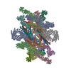
|
|---|---|
| 1 |
|
-Components
-Protein , 9 types, 30 molecules AaBbCEceDFMdfmGJgjHLhlIKikOoNn
| #1: Protein | Mass: 227854.141 Da / Num. of mol.: 2 / Source method: isolated from a natural source / Source: (natural) #2: Protein | Mass: 195848.750 Da / Num. of mol.: 2 / Source method: isolated from a natural source / Source: (natural) #3: Protein | Mass: 93565.156 Da / Num. of mol.: 4 / Source method: isolated from a natural source / Source: (natural) #4: Protein | Mass: 154922.422 Da / Num. of mol.: 6 / Source method: isolated from a natural source / Source: (natural) #5: Protein | Mass: 58618.617 Da / Num. of mol.: 4 / Source method: isolated from a natural source / Source: (natural) #6: Protein | Mass: 55969.496 Da / Num. of mol.: 4 / Source method: isolated from a natural source / Source: (natural) #7: Protein | Mass: 61216.863 Da / Num. of mol.: 4 / Source method: isolated from a natural source / Source: (natural) #8: Protein | Mass: 57120.965 Da / Num. of mol.: 2 / Source method: isolated from a natural source / Source: (natural) #9: Protein | Mass: 74420.305 Da / Num. of mol.: 2 / Source method: isolated from a natural source / Source: (natural) |
|---|
-Experimental details
-Experiment
| Experiment | Method: ELECTRON MICROSCOPY |
|---|---|
| EM experiment | Aggregation state: PARTICLE / 3D reconstruction method: single particle reconstruction |
- Sample preparation
| Component | Name: The IR subunit / Type: COMPLEX / Entity ID: all / Source: NATURAL |
|---|---|
| Source (natural) | Organism: |
| Buffer solution | pH: 7.5 |
| Specimen | Embedding applied: NO / Shadowing applied: NO / Staining applied: NO / Vitrification applied: YES |
| Vitrification | Cryogen name: ETHANE |
- Electron microscopy imaging
| Experimental equipment |  Model: Titan Krios / Image courtesy: FEI Company |
|---|---|
| Microscopy | Model: FEI TITAN KRIOS |
| Electron gun | Electron source:  FIELD EMISSION GUN / Accelerating voltage: 300 kV / Illumination mode: FLOOD BEAM FIELD EMISSION GUN / Accelerating voltage: 300 kV / Illumination mode: FLOOD BEAM |
| Electron lens | Mode: BRIGHT FIELD / Nominal defocus max: 4000 nm / Nominal defocus min: 1200 nm |
| Image recording | Electron dose: 50 e/Å2 / Film or detector model: GATAN K3 (6k x 4k) |
- Processing
| Software | Name: PHENIX / Version: 1.17.1_3660: / Classification: refinement | ||||||||||||||||||||||||
|---|---|---|---|---|---|---|---|---|---|---|---|---|---|---|---|---|---|---|---|---|---|---|---|---|---|
| CTF correction | Type: NONE | ||||||||||||||||||||||||
| 3D reconstruction | Resolution: 4.2 Å / Resolution method: FSC 0.143 CUT-OFF / Num. of particles: 2093631 / Symmetry type: POINT | ||||||||||||||||||||||||
| Refinement | Highest resolution: 4.2 Å | ||||||||||||||||||||||||
| Refine LS restraints |
|
