 Movie
Movie Controller


+ Open data

- Basic information
| Entry | Database: PDB / ID: 7wi7 | |||||||||
|---|---|---|---|---|---|---|---|---|---|---|
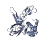
| Title | Crystal structure of human MCM8/9 complex | |||||||||
 Components Components |
| |||||||||
 Keywords Keywords | DNA BINDING PROTEIN / MCM8 / MCM9 / Helicase | |||||||||
| Function / homology |  Function and homology information Function and homology informationMutLbeta complex binding / MutSbeta complex binding / recombinational interstrand cross-link repair / MCM8-MCM9 complex / male gamete generation / mismatch repair involved in maintenance of fidelity involved in DNA-dependent DNA replication / CDC6 association with the ORC:origin complex / female gamete generation / MutSalpha complex binding / E2F-enabled inhibition of pre-replication complex formation ...MutLbeta complex binding / MutSbeta complex binding / recombinational interstrand cross-link repair / MCM8-MCM9 complex / male gamete generation / mismatch repair involved in maintenance of fidelity involved in DNA-dependent DNA replication / CDC6 association with the ORC:origin complex / female gamete generation / MutSalpha complex binding / E2F-enabled inhibition of pre-replication complex formation / Unwinding of DNA / MCM complex / Activation of the pre-replicative complex / Activation of ATR in response to replication stress / protein localization to chromatin / DNA helicase activity / helicase activity / double-strand break repair via homologous recombination / Orc1 removal from chromatin / single-stranded DNA binding / chromosome / DNA helicase / protein stabilization / DNA damage response / chromatin binding / protein-containing complex binding / enzyme binding / ATP hydrolysis activity / nucleoplasm / ATP binding / nucleus Similarity search - Function | |||||||||
| Biological species |  Homo sapiens (human) Homo sapiens (human) | |||||||||
| Method |  X-RAY DIFFRACTION / SYNCHROTRON / MOLECULAR REPLACEMENT / Resolution: 6.6 Å X-RAY DIFFRACTION / SYNCHROTRON / MOLECULAR REPLACEMENT / Resolution: 6.6 Å | |||||||||
 Authors Authors | Li, J. / Liu, Y. | |||||||||
| Funding support |  China, 2items China, 2items
| |||||||||
 Citation Citation | Journal: To Be Published Title: Crystal structure of human MCM8/9 complex Authors: Li, J. / Liu, Y. | |||||||||
| History |
|
- Structure visualization
| Structure viewer | Molecule: MolmilJmol/JSmol |
|---|
- Downloads & links
-Download
| PDBx/mmCIF format | 7wi7.cif.gz | 576.9 KB | Display | PDBx/mmCIF format |
|---|---|---|---|---|
| PDB format | pdb7wi7.ent.gz | Display | PDB format | |
| PDBx/mmJSON format | 7wi7.json.gz | Tree view | PDBx/mmJSON format | |
| Others |  Other downloads Other downloads |
-Validation report
| Summary document | 7wi7_validation.pdf.gz | 572.2 KB | Display | wwPDB validaton report |
|---|---|---|---|---|
| Full document | 7wi7_full_validation.pdf.gz | 585.5 KB | Display | |
| Data in XML | 7wi7_validation.xml.gz | 23.7 KB | Display | |
| Data in CIF | 7wi7_validation.cif.gz | 35.4 KB | Display | |
| Arichive directory | https://data.pdbj.org/pub/pdb/validation_reports/wi/7wi7ftp://data.pdbj.org/pub/pdb/validation_reports/wi/7wi7 | HTTPS FTP |
-Related structure data

-Links
 PDBj
PDBj


- Assembly
| Deposited unit | 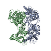
| ||||||||||||
|---|---|---|---|---|---|---|---|---|---|---|---|---|---|
| 1 | 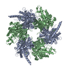
| ||||||||||||
| Unit cell |
|
-Components
| #1: Protein | Mass: 87820.977 Da / Num. of mol.: 1 Source method: isolated from a genetically manipulated source Source: (gene. exp.) Homo sapiens (human) / Gene: MCM8, C20orf154 / Production host:  Trichoplusia ni (cabbage looper) / References: UniProt: Q9UJA3, DNA helicase Trichoplusia ni (cabbage looper) / References: UniProt: Q9UJA3, DNA helicase | ||
|---|---|---|---|
| #2: Protein | Mass: 76848.594 Da / Num. of mol.: 1 Source method: isolated from a genetically manipulated source Source: (gene. exp.) Homo sapiens (human) / Gene: MCM9, C6orf61, MCMDC1 / Production host: Trichoplusia ni (cabbage looper) / References: UniProt: Q9NXL9, DNA helicase | ||
| #3: Chemical |   Mass: 65.409 Da / Num. of mol.: 2 / Source method: obtained synthetically / Formula: Zn / Feature type: SUBJECT OF INVESTIGATION Mass: 65.409 Da / Num. of mol.: 2 / Source method: obtained synthetically / Formula: Zn / Feature type: SUBJECT OF INVESTIGATIONHas ligand of interest | Y | |
-Experimental details
-Experiment
| Experiment | Method: X-RAY DIFFRACTION / Number of used crystals: 1 |
|---|
- Sample preparation
| Crystal | Density Matthews: 3.52 Å3/Da / Density % sol: 65.09 % |
|---|---|
| Crystal grow | Temperature: 289 K / Method: vapor diffusion, hanging drop Details: 0.1M Bis-Tris propone, pH 6.0-7.5, 0.6M sodium citrate |
-Data collection
| Diffraction | Mean temperature: 100 K / Serial crystal experiment: N |
|---|---|
| Diffraction source | Source: SYNCHROTRON / Site: SSRF / Beamline: BL17U / Wavelength: 0.9793 Å |
| Detector | Type: ADSC QUANTUM 315 / Detector: CCD / Date: Nov 11, 2015 |
| Radiation | Protocol: SINGLE WAVELENGTH / Monochromatic (M) / Laue (L): M / Scattering type: x-ray |
| Radiation wavelength | Wavelength: 0.9793 Å / Relative weight: 1 |
| Reflection | Resolution: 6.6→41.5 Å / Num. obs: 4153 / % possible obs: 97.81 % / Redundancy: 13.7 % / Biso Wilson estimate: 431.51 Å2 / CC1/2: 0.99 / Rsym value: 0.061 / Net I/σ(I): 39.8 |
| Reflection shell | Resolution: 6.6→6.71 Å / Redundancy: 14.3 % / Mean I/σ(I) obs: 4.1 / Num. unique obs: 418 / Rsym value: 0.768 / % possible all: 100 |
- Processing
| Software |
| ||||||||||||||||||||||||||||||||||||||||
|---|---|---|---|---|---|---|---|---|---|---|---|---|---|---|---|---|---|---|---|---|---|---|---|---|---|---|---|---|---|---|---|---|---|---|---|---|---|---|---|---|---|
| Refinement | Method to determine structure: MOLECULAR REPLACEMENT Starting model: 7DP3, 7DPD Resolution: 6.6→41.5 Å / SU ML: 1.8622 / Cross valid method: FREE R-VALUE / σ(F): 1.34 / Phase error: 51.0802 Stereochemistry target values: GeoStd + Monomer Library + CDL v1.2
| ||||||||||||||||||||||||||||||||||||||||
| Solvent computation | Shrinkage radii: 0.9 Å / VDW probe radii: 1.11 Å / Solvent model: FLAT BULK SOLVENT MODEL | ||||||||||||||||||||||||||||||||||||||||
| Displacement parameters | Biso mean: 627.19 Å2 | ||||||||||||||||||||||||||||||||||||||||
| Refinement step | Cycle: LAST / Resolution: 6.6→41.5 Å
| ||||||||||||||||||||||||||||||||||||||||
| Refine LS restraints |
| ||||||||||||||||||||||||||||||||||||||||
| LS refinement shell |
| ||||||||||||||||||||||||||||||||||||||||
| Refinement TLS params. | Method: refined / Origin x: 18.1444696396 Å / Origin y: 21.367660008 Å / Origin z: -19.5298083088 Å
| ||||||||||||||||||||||||||||||||||||||||
| Refinement TLS group | Selection details: all |
