 Movie
Movie Controller


+ Open data

- Basic information
| Entry | Database: PDB / ID: 7whf | |||||||||||||||
|---|---|---|---|---|---|---|---|---|---|---|---|---|---|---|---|---|
| Title | Heimdallarchaeota gelsolin (2DGel) bound to rabbit actin | |||||||||||||||
 Components Components |
| |||||||||||||||
 Keywords Keywords | STRUCTURAL PROTEIN / Asgard / gelsolin / actin / filament | |||||||||||||||
| Function / homology |  Function and homology information Function and homology informationactin filament severing / barbed-end actin filament capping / cytoskeletal motor activator activity / myosin heavy chain binding / tropomyosin binding / actin filament bundle / troponin I binding / filamentous actin / mesenchyme migration / skeletal muscle myofibril ...actin filament severing / barbed-end actin filament capping / cytoskeletal motor activator activity / myosin heavy chain binding / tropomyosin binding / actin filament bundle / troponin I binding / filamentous actin / mesenchyme migration / skeletal muscle myofibril / actin filament bundle assembly / striated muscle thin filament / skeletal muscle thin filament assembly / actin monomer binding / skeletal muscle fiber development / stress fiber / titin binding / actin filament polymerization / phosphatidylinositol-4,5-bisphosphate binding / filopodium / actin filament / Hydrolases; Acting on acid anhydrides; Acting on acid anhydrides to facilitate cellular and subcellular movement / calcium-dependent protein binding / actin filament binding / lamellipodium / cell body / protein domain specific binding / hydrolase activity / calcium ion binding / positive regulation of gene expression / magnesium ion binding / ATP binding / metal ion binding / identical protein binding / cytoplasm Similarity search - Function | |||||||||||||||
| Biological species |   Candidatus Heimdallarchaeota archaeon LC_3 (archaea) Candidatus Heimdallarchaeota archaeon LC_3 (archaea) | |||||||||||||||
| Method |  X-RAY DIFFRACTION / SYNCHROTRON / MOLECULAR REPLACEMENT / Resolution: 2.1 Å X-RAY DIFFRACTION / SYNCHROTRON / MOLECULAR REPLACEMENT / Resolution: 2.1 Å | |||||||||||||||
 Authors Authors | Robinson, R.C. / Akil, C. | |||||||||||||||
| Funding support | 4items
| |||||||||||||||
 Citation Citation | Journal: Commun Biol / Year: 2022 Title: Structural and biochemical evidence for the emergence of a calcium-regulated actin cytoskeleton prior to eukaryogenesis Authors: Akil, C. / Tran, L.T. / Orhant-Prioux, M. / Baskaran, Y. / Senju, Y. / Takeda, S. / Chotchuang, P. / Muengsaen, D. / Schulte, A. / Manser, E. / Blanchoin, L. / Robinson, R.C. | |||||||||||||||
| History |
|
- Structure visualization
| Structure viewer | Molecule: MolmilJmol/JSmol |
|---|
- Downloads & links
-Download
| PDBx/mmCIF format | 7whf.cif.gz | 512.2 KB | Display | PDBx/mmCIF format |
|---|---|---|---|---|
| PDB format | pdb7whf.ent.gz | 399.7 KB | Display | PDB format |
| PDBx/mmJSON format | 7whf.json.gz | Tree view | PDBx/mmJSON format | |
| Others |  Other downloads Other downloads |
-Validation report
| Arichive directory | https://data.pdbj.org/pub/pdb/validation_reports/wh/7whfftp://data.pdbj.org/pub/pdb/validation_reports/wh/7whf | HTTPS FTP |
|---|
-Related structure data
| Related structure data | 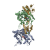 7whgC 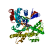 3hbtS S: Starting model for refinement C: citing same article ( |
|---|---|
| Similar structure data |
-Links
 PDBj
PDBj


- Assembly
| Deposited unit | 
| ||||||||||||
|---|---|---|---|---|---|---|---|---|---|---|---|---|---|
| 1 | 
| ||||||||||||
| 2 | 
| ||||||||||||
| Unit cell |
|
-Components
-Protein , 2 types, 4 molecules ABGC
| #1: Protein | Mass: 42109.973 Da / Num. of mol.: 2 Source method: isolated from a genetically manipulated source Source: (gene. exp.) #2: Protein | Mass: 32021.875 Da / Num. of mol.: 2 Source method: isolated from a genetically manipulated source Source: (gene. exp.) Candidatus Heimdallarchaeota archaeon LC_3 (archaea)Gene: HeimC3_12500 / Production host:  |
|---|
-Non-polymers , 4 types, 575 molecules 


| #3: Chemical |  Mass: 507.181 Da / Num. of mol.: 2 / Source method: obtained synthetically / Formula: C10H16N5O13P3 / Comment: ATP, energy-carrying molecule*YM Mass: 507.181 Da / Num. of mol.: 2 / Source method: obtained synthetically / Formula: C10H16N5O13P3 / Comment: ATP, energy-carrying molecule*YM#4: Chemical | ChemComp-CA /  Mass: 40.078 Da / Num. of mol.: 18 / Source method: obtained synthetically / Formula: Ca / Feature type: SUBJECT OF INVESTIGATION Mass: 40.078 Da / Num. of mol.: 18 / Source method: obtained synthetically / Formula: Ca / Feature type: SUBJECT OF INVESTIGATION#5: Chemical |  Mass: 92.094 Da / Num. of mol.: 2 / Source method: obtained synthetically / Formula: C3H8O3 Mass: 92.094 Da / Num. of mol.: 2 / Source method: obtained synthetically / Formula: C3H8O3#6: Water | ChemComp-HOH / | Mass: 18.015 Da / Num. of mol.: 553 / Source method: isolated from a natural source / Formula: H2O |
|---|
-Details
| Has ligand of interest | Y |
|---|
-Experimental details
-Experiment
| Experiment | Method: X-RAY DIFFRACTION / Number of used crystals: 1 |
|---|
- Sample preparation
| Crystal | Density Matthews: 2.22 Å3/Da / Density % sol: 44.52 % |
|---|---|
| Crystal grow | Temperature: 297 K / Method: vapor diffusion, hanging drop Details: 0.4 mM Heimdallarchaeota 2DGel 0.4 mM rabbit actin 1 mM CaCl2 0.1 M HEPES pH 7.0 10% w/v polyethylene glycol 6000 |
-Data collection
| Diffraction | Mean temperature: 100 K / Serial crystal experiment: N |
|---|---|
| Diffraction source | Source: SYNCHROTRON / Site: NSRRC  / Beamline: TPS 05A / Wavelength: 1 Å / Beamline: TPS 05A / Wavelength: 1 Å |
| Detector | Type: RAYONIX MX300-HS / Detector: CCD / Date: May 25, 2018 |
| Radiation | Protocol: SINGLE WAVELENGTH / Monochromatic (M) / Laue (L): M / Scattering type: x-ray |
| Radiation wavelength | Wavelength: 1 Å / Relative weight: 1 |
| Reflection | Resolution: 2.1→20 Å / Num. obs: 63053 / % possible obs: 95.3 % / Redundancy: 5.9 % / CC1/2: 0.954 / Rmerge(I) obs: 0.061 / Rpim(I) all: 0.027 / Rrim(I) all: 0.067 / Net I/σ(I): 24.7 |
| Reflection shell | Resolution: 2.1→2.14 Å / Rmerge(I) obs: 0.651 / Mean I/σ(I) obs: 1.7 / Num. unique obs: 1312 / CC1/2: 0.827 / Rpim(I) all: 0.286 / Rrim(I) all: 0.714 |
- Processing
| Software |
| ||||||||||||||||||||||||
|---|---|---|---|---|---|---|---|---|---|---|---|---|---|---|---|---|---|---|---|---|---|---|---|---|---|
| Refinement | Method to determine structure: MOLECULAR REPLACEMENT Starting model: 3HBT Resolution: 2.1→19.79 Å / Cross valid method: FREE R-VALUE Stereochemistry target values: GeoStd + Monomer Library + CDL v1.2
| ||||||||||||||||||||||||
| Displacement parameters | Biso mean: 39.69 Å2 | ||||||||||||||||||||||||
| Refinement step | Cycle: LAST / Resolution: 2.1→19.79 Å
| ||||||||||||||||||||||||
| Refine LS restraints |
| ||||||||||||||||||||||||
| LS refinement shell | Resolution: 2.1→2.176 Å /
|
