 Movie
Movie Controller

[English] 日本語
 Yorodumi
- PDB-7wgu: Crystal structure of metal-binding protein EfeO from Escherichia coli -
+ Open data

- Basic information
| Entry | Database: PDB / ID: 7wgu | ||||||
|---|---|---|---|---|---|---|---|
| Title | Crystal structure of metal-binding protein EfeO from Escherichia coli | ||||||
 Components Components | Iron uptake system protein EfeO | ||||||
 Keywords Keywords | METAL BINDING PROTEIN / EfeO / Escherichia coli / Iron | ||||||
| Function / homology |  Function and homology information Function and homology informationEfeO/Algp7, imelysin-like domain / : / : / M75 peptidase, HXXE motif / Imelysin-like domain / Imelysin-like domain superfamily / Imelysin / EfeO-type cupredoxin-like domain / Cupredoxin-like domain / A middle domain of Talin 1 ...EfeO/Algp7, imelysin-like domain / : / : / M75 peptidase, HXXE motif / Imelysin-like domain / Imelysin-like domain superfamily / Imelysin / EfeO-type cupredoxin-like domain / Cupredoxin-like domain / A middle domain of Talin 1 / Cupredoxins - blue copper proteins / Cupredoxin / Up-down Bundle / Immunoglobulin-like / Sandwich / Mainly Beta / Mainly Alpha Similarity search - Domain/homology | ||||||
| Biological species |  | ||||||
| Method |  X-RAY DIFFRACTION / SYNCHROTRON / MOLECULAR REPLACEMENT / Resolution: 1.85 Å X-RAY DIFFRACTION / SYNCHROTRON / MOLECULAR REPLACEMENT / Resolution: 1.85 Å | ||||||
 Authors Authors | Nakatsuji, S. / Takase, R. / Mikami, B. / Hashimoto, W. | ||||||
| Funding support |  Japan, 1items Japan, 1items
| ||||||
 Citation Citation | Journal: Biochem.Biophys.Res.Commun. / Year: 2022 Title: Crystal structures of EfeB and EfeO in a bacterial siderophore-independent iron transport system Authors: Nakatsuji, S. / Okumura, K. / Takase, R. / Watanabe, D. / Mikami, B. / Hashimoto, W. | ||||||
| History |
|
- Structure visualization
| Structure viewer | Molecule: MolmilJmol/JSmol |
|---|
- Downloads & links
-Download
| PDBx/mmCIF format | 7wgu.cif.gz | 160.7 KB | Display | PDBx/mmCIF format |
|---|---|---|---|---|
| PDB format | pdb7wgu.ent.gz | 126.8 KB | Display | PDB format |
| PDBx/mmJSON format | 7wgu.json.gz | Tree view | PDBx/mmJSON format | |
| Others |  Other downloads Other downloads |
-Validation report
| Summary document | 7wgu_validation.pdf.gz | 896.7 KB | Display | wwPDB validaton report |
|---|---|---|---|---|
| Full document | 7wgu_full_validation.pdf.gz | 906.1 KB | Display | |
| Data in XML | 7wgu_validation.xml.gz | 30.5 KB | Display | |
| Data in CIF | 7wgu_validation.cif.gz | 44 KB | Display | |
| Arichive directory | https://data.pdbj.org/pub/pdb/validation_reports/wg/7wguftp://data.pdbj.org/pub/pdb/validation_reports/wg/7wgu | HTTPS FTP |
-Related structure data
| Related structure data |  5y4cS S: Starting model for refinement |
|---|---|
| Similar structure data |
-Links
 PDBj
PDBj- Assembly
| Deposited unit | 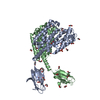
| ||||||||
|---|---|---|---|---|---|---|---|---|---|
| 1 | 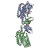
| ||||||||
| Unit cell |
| ||||||||
| Components on special symmetry positions |
|
-Components
-Protein , 1 types, 2 molecules AB
| #1: Protein | Mass: 39557.535 Da / Num. of mol.: 2 Source method: isolated from a genetically manipulated source Source: (gene. exp.) |
|---|
-Non-polymers , 6 types, 356 molecules 


| #2: Chemical | ChemComp-EDO /  Mass: 62.068 Da / Num. of mol.: 30 / Source method: obtained synthetically / Formula: C2H6O2 Mass: 62.068 Da / Num. of mol.: 30 / Source method: obtained synthetically / Formula: C2H6O2#3: Chemical | ChemComp-ACT / |  Mass: 59.044 Da / Num. of mol.: 1 / Source method: obtained synthetically / Formula: C2H3O2 Mass: 59.044 Da / Num. of mol.: 1 / Source method: obtained synthetically / Formula: C2H3O2#4: Chemical | ChemComp-PGE / |  Mass: 150.173 Da / Num. of mol.: 1 / Source method: obtained synthetically / Formula: C6H14O4 Mass: 150.173 Da / Num. of mol.: 1 / Source method: obtained synthetically / Formula: C6H14O4#5: Chemical | ChemComp-SO4 / |  Mass: 96.063 Da / Num. of mol.: 1 / Source method: obtained synthetically / Formula: SO4 Mass: 96.063 Da / Num. of mol.: 1 / Source method: obtained synthetically / Formula: SO4#6: Chemical | ChemComp-ZN / |  Mass: 65.409 Da / Num. of mol.: 1 / Source method: obtained synthetically / Formula: Zn / Feature type: SUBJECT OF INVESTIGATION Mass: 65.409 Da / Num. of mol.: 1 / Source method: obtained synthetically / Formula: Zn / Feature type: SUBJECT OF INVESTIGATION#7: Water | ChemComp-HOH / | Mass: 18.015 Da / Num. of mol.: 322 / Source method: isolated from a natural source / Formula: H2O |
|---|
-Details
| Has ligand of interest | Y |
|---|---|
| Has protein modification | Y |
-Experimental details
-Experiment
| Experiment | Method: X-RAY DIFFRACTION / Number of used crystals: 1 |
|---|
- Sample preparation
| Crystal | Density Matthews: 2.57 Å3/Da / Density % sol: 52.17 % Description: THE ENTRY CONTAINS FRIEDEL PAIRS IN I/F_PLUS/MINUS COLUMNS. |
|---|---|
| Crystal grow | Temperature: 293 K / Method: vapor diffusion, sitting drop / pH: 4.6 Details: 160 mM ammonium sulfate, 80 mM sodium acetate trihydrate (pH 4.6), 20% PEG 4000, 20% glycerol 20% ethylene glycol |
-Data collection
| Diffraction | Mean temperature: 100 K / Serial crystal experiment: N |
|---|---|
| Diffraction source | Source: SYNCHROTRON / Site: SPring-8 / Beamline: BL26B1 / Wavelength: 1 Å |
| Detector | Type: DECTRIS EIGER R 4M / Detector: PIXEL / Date: Feb 13, 2021 |
| Radiation | Protocol: SINGLE WAVELENGTH / Monochromatic (M) / Laue (L): M / Scattering type: x-ray |
| Radiation wavelength | Wavelength: 1 Å / Relative weight: 1 |
| Reflection | Resolution: 1.85→48.15 Å / Num. obs: 65646 / % possible obs: 97.7 % / Redundancy: 3.8 % / Biso Wilson estimate: 37.9 Å2 / CC1/2: 0.999 / Rmerge(I) obs: 0.039 / Rrim(I) all: 0.046 / Net I/σ(I): 18.5 |
| Reflection shell | Resolution: 1.85→1.96 Å / Redundancy: 3.4 % / Rmerge(I) obs: 0.4 / Mean I/σ(I) obs: 2.48 / Num. unique obs: 9994 / CC1/2: 0.918 / Rrim(I) all: 0.469 / % possible all: 92.8 |
- Processing
| Software |
| ||||||||||||||||||||||||||||||||||||||||||||||||||||||||||||||||||||||||||||||||||||||||||||||||||||||||||||||||||||||||||||||||||||||||||||||||||||||||||||||||||||||||
|---|---|---|---|---|---|---|---|---|---|---|---|---|---|---|---|---|---|---|---|---|---|---|---|---|---|---|---|---|---|---|---|---|---|---|---|---|---|---|---|---|---|---|---|---|---|---|---|---|---|---|---|---|---|---|---|---|---|---|---|---|---|---|---|---|---|---|---|---|---|---|---|---|---|---|---|---|---|---|---|---|---|---|---|---|---|---|---|---|---|---|---|---|---|---|---|---|---|---|---|---|---|---|---|---|---|---|---|---|---|---|---|---|---|---|---|---|---|---|---|---|---|---|---|---|---|---|---|---|---|---|---|---|---|---|---|---|---|---|---|---|---|---|---|---|---|---|---|---|---|---|---|---|---|---|---|---|---|---|---|---|---|---|---|---|---|---|---|---|---|
| Refinement | Method to determine structure: MOLECULAR REPLACEMENT Starting model: 5Y4C Resolution: 1.85→48.15 Å / SU ML: 0.22 / Cross valid method: FREE R-VALUE / σ(F): 1.35 / Phase error: 26.98 / Stereochemistry target values: ML
| ||||||||||||||||||||||||||||||||||||||||||||||||||||||||||||||||||||||||||||||||||||||||||||||||||||||||||||||||||||||||||||||||||||||||||||||||||||||||||||||||||||||||
| Solvent computation | Shrinkage radii: 0.9 Å / VDW probe radii: 1.11 Å / Solvent model: FLAT BULK SOLVENT MODEL | ||||||||||||||||||||||||||||||||||||||||||||||||||||||||||||||||||||||||||||||||||||||||||||||||||||||||||||||||||||||||||||||||||||||||||||||||||||||||||||||||||||||||
| Refinement step | Cycle: LAST / Resolution: 1.85→48.15 Å
| ||||||||||||||||||||||||||||||||||||||||||||||||||||||||||||||||||||||||||||||||||||||||||||||||||||||||||||||||||||||||||||||||||||||||||||||||||||||||||||||||||||||||
| Refine LS restraints |
| ||||||||||||||||||||||||||||||||||||||||||||||||||||||||||||||||||||||||||||||||||||||||||||||||||||||||||||||||||||||||||||||||||||||||||||||||||||||||||||||||||||||||
| LS refinement shell |
|
