 Movie
Movie Controller


+ Open data

- Basic information
| Entry | Database: PDB / ID: 7wgi | ||||||
|---|---|---|---|---|---|---|---|
| Title | Crystal structure of AflSQS from Aspergillus flavus | ||||||
 Components Components | Squalene synthase | ||||||
 Keywords Keywords | BIOSYNTHETIC PROTEIN / isoprenoid synthase | ||||||
| Function / homology |  Function and homology information Function and homology informationisoprenoid biosynthetic process via mevalonate / squalene synthase / farnesyl diphosphate metabolic process / squalene synthase [NAD(P)H] activity / ergosterol biosynthetic process / D-glucose transmembrane transporter activity / endoplasmic reticulum membrane Similarity search - Function | ||||||
| Biological species |  | ||||||
| Method |  X-RAY DIFFRACTION / SYNCHROTRON / MOLECULAR REPLACEMENT / Resolution: 2.5 Å X-RAY DIFFRACTION / SYNCHROTRON / MOLECULAR REPLACEMENT / Resolution: 2.5 Å | ||||||
 Authors Authors | Shang, N. / Liu, W.D. / Chen, C.C. / Guo, R.T. | ||||||
| Funding support | 1items
| ||||||
 Citation Citation | Journal: Acs Omega / Year: 2022 Title: A Structural and Bioinformatics Investigation of a Fungal Squalene Synthase and Comparisons with Other Membrane Proteins. Authors: Malwal, S.R. / Shang, N. / Liu, W. / Li, X. / Zhang, L. / Chen, C.C. / Guo, R.T. / Oldfield, E. | ||||||
| History |
|
- Structure visualization
| Structure viewer | Molecule: MolmilJmol/JSmol |
|---|
- Downloads & links
-Download
| PDBx/mmCIF format | 7wgi.cif.gz | 88.4 KB | Display | PDBx/mmCIF format |
|---|---|---|---|---|
| PDB format | pdb7wgi.ent.gz | 65.4 KB | Display | PDB format |
| PDBx/mmJSON format | 7wgi.json.gz | Tree view | PDBx/mmJSON format | |
| Others |  Other downloads Other downloads |
-Validation report
| Arichive directory | https://data.pdbj.org/pub/pdb/validation_reports/wg/7wgiftp://data.pdbj.org/pub/pdb/validation_reports/wg/7wgi | HTTPS FTP |
|---|
-Related structure data
| Related structure data |  7wghC  1ezfS S: Starting model for refinement C: citing same article ( |
|---|---|
| Similar structure data |
-Links
 PDBj
PDBj
- Assembly
| Deposited unit | 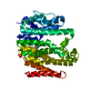
| ||||||||
|---|---|---|---|---|---|---|---|---|---|
| 1 |
| ||||||||
| Unit cell |
|
-Components
| #1: Protein | Mass: 54750.973 Da / Num. of mol.: 1 Source method: isolated from a genetically manipulated source Source: (gene. exp.) Gene: BDV35DRAFT_364291, CA14_004282, F9C07_2287043, G4B11_012149 Plasmid: pET46 / Production host:  |
|---|---|
| #2: Chemical | ChemComp-IND / 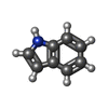  Mass: 117.148 Da / Num. of mol.: 1 / Source method: obtained synthetically / Formula: C8H7N / Feature type: SUBJECT OF INVESTIGATION Mass: 117.148 Da / Num. of mol.: 1 / Source method: obtained synthetically / Formula: C8H7N / Feature type: SUBJECT OF INVESTIGATION |
| #3: Chemical | ChemComp-PO4 /   Mass: 94.971 Da / Num. of mol.: 1 / Source method: obtained synthetically / Formula: PO4 Mass: 94.971 Da / Num. of mol.: 1 / Source method: obtained synthetically / Formula: PO4 |
| #4: Water | ChemComp-HOH /  Mass: 18.015 Da / Num. of mol.: 76 / Source method: isolated from a natural source / Formula: H2O Mass: 18.015 Da / Num. of mol.: 76 / Source method: isolated from a natural source / Formula: H2O |
| Has ligand of interest | Y |
-Experimental details
-Experiment
| Experiment | Method: X-RAY DIFFRACTION / Number of used crystals: 1 |
|---|
- Sample preparation
| Crystal | Density Matthews: 3.35 Å3/Da / Density % sol: 63.27 % |
|---|---|
| Crystal grow | Temperature: 298 K / Method: vapor diffusion, sitting drop / pH: 7 / Details: 1.1 M NaH2PO4, 0.5 M K2HPO4 |
-Data collection
| Diffraction | Mean temperature: 100 K / Serial crystal experiment: N |
|---|---|
| Diffraction source | Source: SYNCHROTRON / Site: NSRRC  / Beamline: BL15A1 / Wavelength: 0.9998 Å / Beamline: BL15A1 / Wavelength: 0.9998 Å |
| Detector | Type: RAYONIX MX-300 / Detector: CCD / Date: Oct 10, 2019 |
| Radiation | Protocol: SINGLE WAVELENGTH / Monochromatic (M) / Laue (L): M / Scattering type: x-ray |
| Radiation wavelength | Wavelength: 0.9998 Å / Relative weight: 1 |
| Reflection | Resolution: 2.5→25 Å / Num. obs: 25222 / % possible obs: 100 % / Redundancy: 6.9 % / CC1/2: 0.913 / Net I/σ(I): 31.05 |
| Reflection shell | Resolution: 2.5→2.59 Å / Redundancy: 6.9 % / Mean I/σ(I) obs: 2.18 / Num. unique obs: 2508 / CC1/2: 0.626 / % possible all: 100 |
- Processing
| Software |
| ||||||||||||||||||||||||||||||||||||||||||||||||||||||||||||
|---|---|---|---|---|---|---|---|---|---|---|---|---|---|---|---|---|---|---|---|---|---|---|---|---|---|---|---|---|---|---|---|---|---|---|---|---|---|---|---|---|---|---|---|---|---|---|---|---|---|---|---|---|---|---|---|---|---|---|---|---|---|
| Refinement | Method to determine structure: MOLECULAR REPLACEMENT Starting model: 1EZF Resolution: 2.5→24.33 Å / SU ML: 0.32 / Cross valid method: THROUGHOUT / σ(F): 1.34 / Phase error: 25.76 / Stereochemistry target values: ML
| ||||||||||||||||||||||||||||||||||||||||||||||||||||||||||||
| Solvent computation | Shrinkage radii: 0.9 Å / VDW probe radii: 1.11 Å / Solvent model: FLAT BULK SOLVENT MODEL | ||||||||||||||||||||||||||||||||||||||||||||||||||||||||||||
| Displacement parameters | Biso max: 132.72 Å2 / Biso mean: 63.68 Å2 / Biso min: 30 Å2 | ||||||||||||||||||||||||||||||||||||||||||||||||||||||||||||
| Refinement step | Cycle: final / Resolution: 2.5→24.33 Å
| ||||||||||||||||||||||||||||||||||||||||||||||||||||||||||||
| LS refinement shell | Refine-ID: X-RAY DIFFRACTION / Rfactor Rfree error: 0 / Total num. of bins used: 9 / % reflection obs: 100 %
|
