 Movie
Movie Controller

[English] 日本語
 Yorodumi
- PDB-7wf8: Crystal structure of mouse SNX25 RGS domain in space group P212121 -
+ Open data

- Basic information
| Entry | Database: PDB / ID: 7wf8 | ||||||
|---|---|---|---|---|---|---|---|
| Title | Crystal structure of mouse SNX25 RGS domain in space group P212121 | ||||||
 Components Components | Sorting nexin-25 | ||||||
 Keywords Keywords | PROTEIN TRANSPORT / Sorting nexin / SNX25 / RGS | ||||||
| Function / homology |  Function and homology information Function and homology informationreceptor catabolic process / type I transforming growth factor beta receptor binding / phosphatidylinositol binding / negative regulation of transforming growth factor beta receptor signaling pathway / protein transport / endosome membrane / endosome Similarity search - Function | ||||||
| Biological species |  | ||||||
| Method |  X-RAY DIFFRACTION / SYNCHROTRON / MOLECULAR REPLACEMENT / Resolution: 1.35 Å X-RAY DIFFRACTION / SYNCHROTRON / MOLECULAR REPLACEMENT / Resolution: 1.35 Å | ||||||
 Authors Authors | Zhang, Y. / Xu, J. / Liu, J. | ||||||
| Funding support |  China, 1items China, 1items
| ||||||
 Citation Citation | Journal: J.Mol.Biol. / Year: 2022 Title: Structural Studies Reveal Unique Non-canonical Regulators of G Protein Signaling Homology (RH) Domains in Sorting Nexins. Authors: Zhang, Y. / Chen, R. / Dong, Y. / Zhu, J. / Su, K. / Liu, J. / Xu, J. | ||||||
| History |
|
- Structure visualization
| Structure viewer | Molecule: MolmilJmol/JSmol |
|---|
- Downloads & links
-Download
| PDBx/mmCIF format | 7wf8.cif.gz | 78.6 KB | Display | PDBx/mmCIF format |
|---|---|---|---|---|
| PDB format | pdb7wf8.ent.gz | 57.2 KB | Display | PDB format |
| PDBx/mmJSON format | 7wf8.json.gz | Tree view | PDBx/mmJSON format | |
| Others |  Other downloads Other downloads |
-Validation report
| Summary document | 7wf8_validation.pdf.gz | 3.3 MB | Display | wwPDB validaton report |
|---|---|---|---|---|
| Full document | 7wf8_full_validation.pdf.gz | 3.3 MB | Display | |
| Data in XML | 7wf8_validation.xml.gz | 16 KB | Display | |
| Data in CIF | 7wf8_validation.cif.gz | 23.1 KB | Display | |
| Arichive directory | https://data.pdbj.org/pub/pdb/validation_reports/wf/7wf8ftp://data.pdbj.org/pub/pdb/validation_reports/wf/7wf8 | HTTPS FTP |
-Related structure data
| Related structure data | 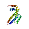 7wf6C 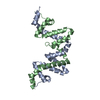 7wf9C 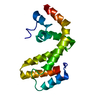 1zv4S S: Starting model for refinement C: citing same article ( |
|---|---|
| Similar structure data |
-Links
 PDBj
PDBj
- Assembly
| Deposited unit | 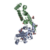
| ||||||||
|---|---|---|---|---|---|---|---|---|---|
| 1 | 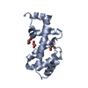
| ||||||||
| 2 | 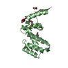
| ||||||||
| Unit cell |
|
-Components
| #1: Protein | Mass: 15763.890 Da / Num. of mol.: 2 Source method: isolated from a genetically manipulated source Source: (gene. exp.)  #2: Chemical | ChemComp-GOL /   Mass: 92.094 Da / Num. of mol.: 4 / Source method: obtained synthetically / Formula: C3H8O3 / Feature type: SUBJECT OF INVESTIGATION Mass: 92.094 Da / Num. of mol.: 4 / Source method: obtained synthetically / Formula: C3H8O3 / Feature type: SUBJECT OF INVESTIGATION#3: Chemical | ChemComp-PEG /   Mass: 106.120 Da / Num. of mol.: 7 / Source method: obtained synthetically / Formula: C4H10O3 / Feature type: SUBJECT OF INVESTIGATION Mass: 106.120 Da / Num. of mol.: 7 / Source method: obtained synthetically / Formula: C4H10O3 / Feature type: SUBJECT OF INVESTIGATION#4: Chemical | ChemComp-CL / |   Mass: 35.453 Da / Num. of mol.: 1 / Source method: obtained synthetically / Formula: Cl / Feature type: SUBJECT OF INVESTIGATION Mass: 35.453 Da / Num. of mol.: 1 / Source method: obtained synthetically / Formula: Cl / Feature type: SUBJECT OF INVESTIGATION#5: Water | ChemComp-HOH / |  Mass: 18.015 Da / Num. of mol.: 277 / Source method: isolated from a natural source / Formula: H2O Mass: 18.015 Da / Num. of mol.: 277 / Source method: isolated from a natural source / Formula: H2OHas ligand of interest | Y | |
|---|
-Experimental details
-Experiment
| Experiment | Method: X-RAY DIFFRACTION / Number of used crystals: 1 |
|---|
- Sample preparation
| Crystal | Density Matthews: 2.42 Å3/Da / Density % sol: 49.26 % |
|---|---|
| Crystal grow | Temperature: 277 K / Method: vapor diffusion / pH: 8.5 / Details: 0.1M Tris pH 8.5, 25% PEG 8000 |
-Data collection
| Diffraction | Mean temperature: 100 K / Serial crystal experiment: N | |||||||||||||||||||||||||||
|---|---|---|---|---|---|---|---|---|---|---|---|---|---|---|---|---|---|---|---|---|---|---|---|---|---|---|---|---|
| Diffraction source | Source: SYNCHROTRON / Site: SSRF / Beamline: BL19U1 / Wavelength: 0.9778 Å | |||||||||||||||||||||||||||
| Detector | Type: DECTRIS PILATUS3 6M / Detector: PIXEL / Date: Jun 1, 2017 | |||||||||||||||||||||||||||
| Radiation | Protocol: SINGLE WAVELENGTH / Monochromatic (M) / Laue (L): M / Scattering type: x-ray | |||||||||||||||||||||||||||
| Radiation wavelength | Wavelength: 0.9778 Å / Relative weight: 1 | |||||||||||||||||||||||||||
| Reflection | Resolution: 1.35→52.42 Å / Num. obs: 68289 / % possible obs: 99.9 % / Redundancy: 8.1 % / CC1/2: 0.999 / Rmerge(I) obs: 0.069 / Rpim(I) all: 0.025 / Rrim(I) all: 0.073 / Net I/σ(I): 13.5 / Num. measured all: 553629 / Scaling rejects: 367 | |||||||||||||||||||||||||||
| Reflection shell | Diffraction-ID: 1 / % possible all: 99.9
|
- Processing
| Software |
| ||||||||||||||||||||||||||||||||||||||||||||||||||||||||||||
|---|---|---|---|---|---|---|---|---|---|---|---|---|---|---|---|---|---|---|---|---|---|---|---|---|---|---|---|---|---|---|---|---|---|---|---|---|---|---|---|---|---|---|---|---|---|---|---|---|---|---|---|---|---|---|---|---|---|---|---|---|---|
| Refinement | Method to determine structure: MOLECULAR REPLACEMENT Starting model: 1ZV4 Resolution: 1.35→48.99 Å / Cor.coef. Fo:Fc: 0.973 / Cor.coef. Fo:Fc free: 0.963 / SU B: 0.899 / SU ML: 0.036 / SU R Cruickshank DPI: 0.0489 / Cross valid method: THROUGHOUT / σ(F): 0 / ESU R: 0.049 / ESU R Free: 0.052 / Stereochemistry target values: MAXIMUM LIKELIHOOD Details: HYDROGENS HAVE BEEN ADDED IN THE RIDING POSITIONS U VALUES : REFINED INDIVIDUALLY
| ||||||||||||||||||||||||||||||||||||||||||||||||||||||||||||
| Solvent computation | Ion probe radii: 0.8 Å / Shrinkage radii: 0.8 Å / VDW probe radii: 1.2 Å / Solvent model: MASK | ||||||||||||||||||||||||||||||||||||||||||||||||||||||||||||
| Displacement parameters | Biso max: 69.96 Å2 / Biso mean: 19.504 Å2 / Biso min: 10.46 Å2
| ||||||||||||||||||||||||||||||||||||||||||||||||||||||||||||
| Refinement step | Cycle: final / Resolution: 1.35→48.99 Å
| ||||||||||||||||||||||||||||||||||||||||||||||||||||||||||||
| Refine LS restraints |
| ||||||||||||||||||||||||||||||||||||||||||||||||||||||||||||
| LS refinement shell | Resolution: 1.35→1.385 Å / Rfactor Rfree error: 0 / Total num. of bins used: 20
|
