[English] 日本語
 Yorodumi
- PDB-7wek: Crystal structure of the mouse Wdr47 NTD in complex with the WBR ... -
+ Open data
- Basic information
| Entry | Database: PDB / ID: 7wek | ||||||
|---|---|---|---|---|---|---|---|
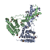
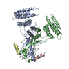
| Title | Crystal structure of the mouse Wdr47 NTD in complex with the WBR motif form Camsap3. | ||||||
 Components Components |
| ||||||
 Keywords Keywords | PROTEIN BINDING / LisH motif containing protein | ||||||
| Function / homology |  Function and homology information Function and homology informationregulation of organelle organization / detection of hot stimulus involved in thermoception / zonula adherens maintenance / anterior commissure morphogenesis / microtubule minus-end / cerebral cortex radial glia-guided migration / protein transport along microtubule / microtubule anchoring / regulation of Golgi organization / microtubule minus-end binding ...regulation of organelle organization / detection of hot stimulus involved in thermoception / zonula adherens maintenance / anterior commissure morphogenesis / microtubule minus-end / cerebral cortex radial glia-guided migration / protein transport along microtubule / microtubule anchoring / regulation of Golgi organization / microtubule minus-end binding / establishment or maintenance of microtubule cytoskeleton polarity / cilium movement / epithelial cell-cell adhesion / zonula adherens / corpus callosum development / neuronal stem cell population maintenance / negative regulation of microtubule depolymerization / establishment of epithelial cell apical/basal polarity / embryo development ending in birth or egg hatching / neural precursor cell proliferation / regulation of focal adhesion assembly / motile cilium / spectrin binding / motor behavior / regulation of microtubule polymerization / axoneme / regulation of microtubule cytoskeleton organization / regulation of cell migration / adult locomotory behavior / locomotory behavior / brain development / cerebral cortex development / autophagy / microtubule cytoskeleton organization / neuron projection development / actin filament binding / microtubule cytoskeleton / growth cone / in utero embryonic development / microtubule / calmodulin binding / neuron projection / ciliary basal body / axon / neuronal cell body / dendrite / centrosome / protein-containing complex binding / cytoplasm Similarity search - Function | ||||||
| Biological species |  | ||||||
| Method |  X-RAY DIFFRACTION / X-RAY DIFFRACTION /  SYNCHROTRON / SYNCHROTRON /  MOLECULAR REPLACEMENT / Resolution: 3.21 Å MOLECULAR REPLACEMENT / Resolution: 3.21 Å | ||||||
 Authors Authors | Ren, J.Q. / Li, D. / Feng, W. | ||||||
| Funding support |  China, 1items China, 1items
| ||||||
 Citation Citation |  Journal: Cell Rep / Year: 2022 Journal: Cell Rep / Year: 2022Title: Intertwined Wdr47-NTD dimer recognizes a basic-helical motif in Camsap proteins for proper central-pair microtubule formation. Authors: Ren, J. / Li, D. / Liu, J. / Liu, H. / Yan, X. / Zhu, X. / Feng, W. | ||||||
| History |
|
- Structure visualization
| Structure viewer | Molecule:  Molmil Molmil Jmol/JSmol Jmol/JSmol |
|---|
- Downloads & links
- Download
| PDBx/mmCIF format |  7wek.cif.gz 7wek.cif.gz | 118.4 KB | Display |  PDBx/mmCIF format PDBx/mmCIF format |
|---|---|---|---|---|
| PDB format |  pdb7wek.ent.gz pdb7wek.ent.gz | 90.8 KB | Display |  PDB format PDB format |
| PDBx/mmJSON format |  7wek.json.gz 7wek.json.gz | Tree view |  PDBx/mmJSON format PDBx/mmJSON format | |
| Others |  Other downloads Other downloads |
-Validation report
| Summary document |  7wek_validation.pdf.gz 7wek_validation.pdf.gz | 446.1 KB | Display |  wwPDB validaton report wwPDB validaton report |
|---|---|---|---|---|
| Full document |  7wek_full_validation.pdf.gz 7wek_full_validation.pdf.gz | 448.5 KB | Display | |
| Data in XML |  7wek_validation.xml.gz 7wek_validation.xml.gz | 19.9 KB | Display | |
| Data in CIF |  7wek_validation.cif.gz 7wek_validation.cif.gz | 26.2 KB | Display | |
| Arichive directory |  https://data.pdbj.org/pub/pdb/validation_reports/we/7wek https://data.pdbj.org/pub/pdb/validation_reports/we/7wek ftp://data.pdbj.org/pub/pdb/validation_reports/we/7wek ftp://data.pdbj.org/pub/pdb/validation_reports/we/7wek | HTTPS FTP |
-Related structure data
| Related structure data |  7wejSC S: Starting model for refinement C: citing same article ( |
|---|---|
| Similar structure data | Similarity search - Function & homology  F&H Search F&H Search |
- Links
Links
- Assembly
| Deposited unit | 
| ||||||||
|---|---|---|---|---|---|---|---|---|---|
| 1 |
| ||||||||
| Unit cell |
|
- Components
| #1: Protein | Mass: 33550.934 Da / Num. of mol.: 2 Source method: isolated from a genetically manipulated source Source: (gene. exp.)   #2: Protein/peptide | Mass: 3215.610 Da / Num. of mol.: 2 / Source method: obtained synthetically / Source: (synth.)  |
|---|
-Experimental details
-Experiment
| Experiment | Method:  X-RAY DIFFRACTION / Number of used crystals: 1 X-RAY DIFFRACTION / Number of used crystals: 1 |
|---|
- Sample preparation
| Crystal | Density Matthews: 3.24 Å3/Da / Density % sol: 62.05 % |
|---|---|
| Crystal grow | Temperature: 289 K / Method: vapor diffusion, sitting drop / pH: 8.5 / Details: 0.1M Tris-HCl, 20% (w/v) PEG 3350 |
-Data collection
| Diffraction | Mean temperature: 100 K / Serial crystal experiment: N |
|---|---|
| Diffraction source | Source:  SYNCHROTRON / Site: SYNCHROTRON / Site:  SSRF SSRF  / Beamline: BL17U / Wavelength: 0.979 Å / Beamline: BL17U / Wavelength: 0.979 Å |
| Detector | Type: DECTRIS EIGER X 16M / Detector: PIXEL / Date: Jul 19, 2020 |
| Radiation | Protocol: SINGLE WAVELENGTH / Monochromatic (M) / Laue (L): M / Scattering type: x-ray |
| Radiation wavelength | Wavelength: 0.979 Å / Relative weight: 1 |
| Reflection | Resolution: 3.2→30 Å / Num. obs: 16169 / % possible obs: 99.8 % / Redundancy: 6.7 % / CC1/2: 0.994 / CC star: 0.999 / Net I/σ(I): 18.3 |
| Reflection shell | Resolution: 3.2→3.31 Å / Mean I/σ(I) obs: 1.148 / Num. unique obs: 1569 / CC1/2: 0.664 / CC star: 0.893 |
- Processing
| Software |
| ||||||||||||||||||||||||||||||||||||||||||||||||||||||||||||
|---|---|---|---|---|---|---|---|---|---|---|---|---|---|---|---|---|---|---|---|---|---|---|---|---|---|---|---|---|---|---|---|---|---|---|---|---|---|---|---|---|---|---|---|---|---|---|---|---|---|---|---|---|---|---|---|---|---|---|---|---|---|
| Refinement | Method to determine structure:  MOLECULAR REPLACEMENT MOLECULAR REPLACEMENTStarting model: 7WEJ Resolution: 3.21→24.18 Å / SU ML: 0.48 / Cross valid method: THROUGHOUT / σ(F): 1.36 / Phase error: 34.42 / Stereochemistry target values: ML
| ||||||||||||||||||||||||||||||||||||||||||||||||||||||||||||
| Solvent computation | Shrinkage radii: 0.9 Å / VDW probe radii: 1.11 Å / Solvent model: FLAT BULK SOLVENT MODEL | ||||||||||||||||||||||||||||||||||||||||||||||||||||||||||||
| Displacement parameters | Biso max: 240.53 Å2 / Biso min: 28.94 Å2 | ||||||||||||||||||||||||||||||||||||||||||||||||||||||||||||
| Refinement step | Cycle: final / Resolution: 3.21→24.18 Å
| ||||||||||||||||||||||||||||||||||||||||||||||||||||||||||||
| LS refinement shell | Refine-ID: X-RAY DIFFRACTION / Rfactor Rfree error: 0
|
 Movie
Movie Controller


 PDBj
PDBj
