 Movie
Movie Controller

[English] 日本語
 Yorodumi
- PDB-7wdr: Crystal structures of MeBglD2 in complex with various saccharides -
+ Open data

- Basic information
| Entry | Database: PDB / ID: 7wdr | ||||||
|---|---|---|---|---|---|---|---|
| Title | Crystal structures of MeBglD2 in complex with various saccharides | ||||||
 Components Components | Beta-glucosidase | ||||||
 Keywords Keywords | HYDROLASE / glycoside hydrolase family 1 | ||||||
| Function / homology |  Function and homology information Function and homology informationbeta-glucosidase / beta-glucosidase activity / cellulose catabolic process Similarity search - Function | ||||||
| Biological species | metagenome (others) | ||||||
| Method |  X-RAY DIFFRACTION / SYNCHROTRON / MOLECULAR REPLACEMENT / Resolution: 2 Å X-RAY DIFFRACTION / SYNCHROTRON / MOLECULAR REPLACEMENT / Resolution: 2 Å | ||||||
 Authors Authors | Watanabe, M. / Matsuzawa, T. / Nakamichi, Y. / Akita, H. / Yaoi, K. | ||||||
| Funding support |  Japan, 1items Japan, 1items
| ||||||
 Citation Citation | Journal: Appl.Microbiol.Biotechnol. / Year: 2022 Title: Crystal structure of metagenomic beta-glycosidase MeBglD2 in complex with various saccharides Authors: Matsuzawa, T. / Watanabe, M. / Nakamichi, Y. / Akita, H. / Yaoi, K. | ||||||
| History |
|
- Structure visualization
| Structure viewer | Molecule: MolmilJmol/JSmol |
|---|
- Downloads & links
-Download
| PDBx/mmCIF format | 7wdr.cif.gz | 106.6 KB | Display | PDBx/mmCIF format |
|---|---|---|---|---|
| PDB format | pdb7wdr.ent.gz | 80.8 KB | Display | PDB format |
| PDBx/mmJSON format | 7wdr.json.gz | Tree view | PDBx/mmJSON format | |
| Others |  Other downloads Other downloads |
-Validation report
| Arichive directory | https://data.pdbj.org/pub/pdb/validation_reports/wd/7wdrftp://data.pdbj.org/pub/pdb/validation_reports/wd/7wdr | HTTPS FTP |
|---|
-Related structure data
| Related structure data |  7wdoC  7wdpC  7wdsC  7wdvC  5xgzS S: Starting model for refinement C: citing same article ( |
|---|---|
| Similar structure data |
-Links
 PDBj
PDBj- Assembly
| Deposited unit | 
| ||||||||
|---|---|---|---|---|---|---|---|---|---|
| 1 |
| ||||||||
| Unit cell |
|
-Components
| #1: Protein | Mass: 52127.074 Da / Num. of mol.: 1 / Mutation: E356A Source method: isolated from a genetically manipulated source Source: (gene. exp.) metagenome (others) / Production host:  | ||||||
|---|---|---|---|---|---|---|---|
| #2: Sugar | 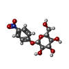  Type: D-saccharide / Mass: 301.249 Da / Num. of mol.: 2 Type: D-saccharide / Mass: 301.249 Da / Num. of mol.: 2Source method: isolated from a genetically manipulated source Formula: C12H15NO8 / Feature type: SUBJECT OF INVESTIGATION #3: Chemical |   Mass: 96.063 Da / Num. of mol.: 3 / Source method: obtained synthetically / Formula: SO4 / Feature type: SUBJECT OF INVESTIGATION Mass: 96.063 Da / Num. of mol.: 3 / Source method: obtained synthetically / Formula: SO4 / Feature type: SUBJECT OF INVESTIGATION#4: Water | ChemComp-HOH / |  Mass: 18.015 Da / Num. of mol.: 41 / Source method: isolated from a natural source / Formula: H2O Mass: 18.015 Da / Num. of mol.: 41 / Source method: isolated from a natural source / Formula: H2OHas ligand of interest | Y | |
-Experimental details
-Experiment
| Experiment | Method: X-RAY DIFFRACTION / Number of used crystals: 1 |
|---|
- Sample preparation
| Crystal | Density Matthews: 3.28 Å3/Da / Density % sol: 62.53 % |
|---|---|
| Crystal grow | Temperature: 293 K / Method: vapor diffusion, hanging drop Details: 0.1 M Tris-HCl buffer (pH 8.5), 1.2 M lithium sulfate, 0.01 M nickel chloride |
-Data collection
| Diffraction | Mean temperature: 100 K / Serial crystal experiment: N |
|---|---|
| Diffraction source | Source: SYNCHROTRON / Site: SPring-8 / Beamline: BL44XU / Wavelength: 0.9 Å |
| Detector | Type: RAYONIX MX300HE / Detector: CCD / Date: Oct 5, 2016 |
| Radiation | Protocol: SINGLE WAVELENGTH / Monochromatic (M) / Laue (L): M / Scattering type: x-ray |
| Radiation wavelength | Wavelength: 0.9 Å / Relative weight: 1 |
| Reflection | Resolution: 2→50 Å / Num. obs: 46907 / % possible obs: 99.5 % / Redundancy: 12.2 % / Rmerge(I) obs: 0.136 / Net I/σ(I): 10.5 |
| Reflection shell | Resolution: 2→2.03 Å / Rmerge(I) obs: 0.777 / Num. unique obs: 2294 |
- Processing
| Software |
| ||||||||||||||||||||||||||||||||||||||||||||||||||||||||||||||||||||||||||||||||||||||||||||||||||||||||||||||||||||||||||||||||||||||||||||||||||||||||||||||||||||||||||||||||||||||
|---|---|---|---|---|---|---|---|---|---|---|---|---|---|---|---|---|---|---|---|---|---|---|---|---|---|---|---|---|---|---|---|---|---|---|---|---|---|---|---|---|---|---|---|---|---|---|---|---|---|---|---|---|---|---|---|---|---|---|---|---|---|---|---|---|---|---|---|---|---|---|---|---|---|---|---|---|---|---|---|---|---|---|---|---|---|---|---|---|---|---|---|---|---|---|---|---|---|---|---|---|---|---|---|---|---|---|---|---|---|---|---|---|---|---|---|---|---|---|---|---|---|---|---|---|---|---|---|---|---|---|---|---|---|---|---|---|---|---|---|---|---|---|---|---|---|---|---|---|---|---|---|---|---|---|---|---|---|---|---|---|---|---|---|---|---|---|---|---|---|---|---|---|---|---|---|---|---|---|---|---|---|---|---|
| Refinement | Method to determine structure: MOLECULAR REPLACEMENT Starting model: 5XGZ Resolution: 2→19.99 Å / Cor.coef. Fo:Fc: 0.975 / Cor.coef. Fo:Fc free: 0.955 / SU B: 5.447 / SU ML: 0.136 / Cross valid method: THROUGHOUT / ESU R: 0.134 / ESU R Free: 0.143 Stereochemistry target values: MAXIMUM LIKELIHOOD WITH PHASES Details: HYDROGENS HAVE BEEN ADDED IN THE RIDING POSITIONS
| ||||||||||||||||||||||||||||||||||||||||||||||||||||||||||||||||||||||||||||||||||||||||||||||||||||||||||||||||||||||||||||||||||||||||||||||||||||||||||||||||||||||||||||||||||||||
| Solvent computation | Ion probe radii: 0.8 Å / Shrinkage radii: 0.8 Å / VDW probe radii: 1.2 Å / Solvent model: MASK | ||||||||||||||||||||||||||||||||||||||||||||||||||||||||||||||||||||||||||||||||||||||||||||||||||||||||||||||||||||||||||||||||||||||||||||||||||||||||||||||||||||||||||||||||||||||
| Displacement parameters | Biso mean: 56.606 Å2
| ||||||||||||||||||||||||||||||||||||||||||||||||||||||||||||||||||||||||||||||||||||||||||||||||||||||||||||||||||||||||||||||||||||||||||||||||||||||||||||||||||||||||||||||||||||||
| Refinement step | Cycle: 1 / Resolution: 2→19.99 Å
| ||||||||||||||||||||||||||||||||||||||||||||||||||||||||||||||||||||||||||||||||||||||||||||||||||||||||||||||||||||||||||||||||||||||||||||||||||||||||||||||||||||||||||||||||||||||
| Refine LS restraints |
|