 Movie
Movie Controller

[English] 日本語
 Yorodumi
- PDB-7wda: Crystal structure LpqY in complex with Trehalose from Mycobacteri... -
+ Open data

- Basic information
| Entry | Database: PDB / ID: 7wda | ||||||
|---|---|---|---|---|---|---|---|
| Title | Crystal structure LpqY in complex with Trehalose from Mycobacterium tuberculosis | ||||||
 Components Components | Trehalose-binding lipoprotein LpqY | ||||||
 Keywords Keywords | SUGAR BINDING PROTEIN / Mycobacterium tuberculosis / LpqY / Trehalose / SugABC | ||||||
| Function / homology |  Function and homology information Function and homology informationtrehalose transmembrane transporter activity / trehalose transport / biological process involved in interaction with host / ATP-binding cassette (ABC) transporter complex / periplasmic space / plasma membrane Similarity search - Function | ||||||
| Biological species |  Mycobacterium tuberculosis H37Rv (bacteria) Mycobacterium tuberculosis H37Rv (bacteria) | ||||||
| Method |  X-RAY DIFFRACTION / SYNCHROTRON / MOLECULAR REPLACEMENT / Resolution: 1.91 Å X-RAY DIFFRACTION / SYNCHROTRON / MOLECULAR REPLACEMENT / Resolution: 1.91 Å | ||||||
 Authors Authors | Sharma, D. / Das, U. | ||||||
| Funding support |  India, 1items India, 1items
| ||||||
 Citation Citation | Journal: Acta Crystallogr D Struct Biol / Year: 2022 Title: Structural analysis of LpqY, a substrate-binding protein from the SugABC transporter of Mycobacterium tuberculosis, provides insights into its trehalose specificity. Authors: Sharma, D. / Singh, M. / Kaur, P. / Das, U. | ||||||
| History |
|
- Structure visualization
| Structure viewer | Molecule: MolmilJmol/JSmol |
|---|
- Downloads & links
-Download
| PDBx/mmCIF format | 7wda.cif.gz | 738.6 KB | Display | PDBx/mmCIF format |
|---|---|---|---|---|
| PDB format | pdb7wda.ent.gz | 544.5 KB | Display | PDB format |
| PDBx/mmJSON format | 7wda.json.gz | Tree view | PDBx/mmJSON format | |
| Others |  Other downloads Other downloads |
-Validation report
| Summary document | 7wda_validation.pdf.gz | 2.1 MB | Display | wwPDB validaton report |
|---|---|---|---|---|
| Full document | 7wda_full_validation.pdf.gz | 2.1 MB | Display | |
| Data in XML | 7wda_validation.xml.gz | 65.8 KB | Display | |
| Data in CIF | 7wda_validation.cif.gz | 96.2 KB | Display | |
| Arichive directory | https://data.pdbj.org/pub/pdb/validation_reports/wd/7wdaftp://data.pdbj.org/pub/pdb/validation_reports/wd/7wda | HTTPS FTP |
-Related structure data
| Related structure data |  7wcjSC S: Starting model for refinement C: citing same article ( |
|---|---|
| Similar structure data |
-Links
 PDBj
PDBj


- Assembly
| Deposited unit | 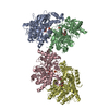
| ||||||||||
|---|---|---|---|---|---|---|---|---|---|---|---|
| 1 | 
| ||||||||||
| 2 | 
| ||||||||||
| 3 | 
| ||||||||||
| 4 | 
| ||||||||||
| Unit cell |
|
-Components
| #1: Protein | Mass: 47353.098 Da / Num. of mol.: 4 Source method: isolated from a genetically manipulated source Source: (gene. exp.) Mycobacterium tuberculosis H37Rv (bacteria)Gene: lpqY, Rv1235 / Plasmid: pET / Production host: #2: Polysaccharide | alpha-D-glucopyranose-(1-1)-alpha-D-glucopyranose 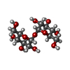  Source method: isolated from a genetically manipulated source Details: oligosaccharide with reducing-end-to-reducing-end glycosidic bond References: trehalose #3: Chemical | ChemComp-SO4 /   Mass: 96.063 Da / Num. of mol.: 4 / Source method: obtained synthetically / Formula: SO4 Mass: 96.063 Da / Num. of mol.: 4 / Source method: obtained synthetically / Formula: SO4#4: Water | ChemComp-HOH / |  Mass: 18.015 Da / Num. of mol.: 941 / Source method: isolated from a natural source / Formula: H2O Mass: 18.015 Da / Num. of mol.: 941 / Source method: isolated from a natural source / Formula: H2OHas ligand of interest | Y | Has protein modification | Y | |
|---|
-Experimental details
-Experiment
| Experiment | Method: X-RAY DIFFRACTION / Number of used crystals: 1 |
|---|
- Sample preparation
| Crystal | Density Matthews: 2.36 Å3/Da / Density % sol: 47.83 % |
|---|---|
| Crystal grow | Temperature: 289 K / Method: vapor diffusion, sitting drop / Details: PEG |
-Data collection
| Diffraction | Mean temperature: 100 K / Serial crystal experiment: N |
|---|---|
| Diffraction source | Source: SYNCHROTRON / Site: ESRF  / Beamline: ID30B / Wavelength: 0.9763 Å / Beamline: ID30B / Wavelength: 0.9763 Å |
| Detector | Type: DECTRIS PILATUS 6M / Detector: PIXEL / Date: Nov 26, 2021 / Details: Vertical CRL / Horizontal Eliptical mirror |
| Radiation | Monochromator: Si(111) / Protocol: SINGLE WAVELENGTH / Monochromatic (M) / Laue (L): M / Scattering type: x-ray |
| Radiation wavelength | Wavelength: 0.9763 Å / Relative weight: 1 |
| Reflection | Resolution: 1.91→48.03 Å / Num. obs: 137970 / % possible obs: 99.52 % / Redundancy: 12.9 % / Biso Wilson estimate: 33.12 Å2 / CC1/2: 0.998 / CC star: 0.99 / Rmerge(I) obs: 0.24 / Rpim(I) all: 0.07 / Net I/σ(I): 8.43 |
| Reflection shell | Resolution: 1.91→1.97 Å / Redundancy: 11.6 % / Rmerge(I) obs: 4.29 / Mean I/σ(I) obs: 0.54 / Num. unique obs: 13670 / CC1/2: 0.279 / Rpim(I) all: 1.3 / % possible all: 95.83 |
- Processing
| Software |
| |||||||||||||||||||||||||||||||||||||||||||||||||||||||||||||||||||||||||||||||||||||||||||||||||||||||||||||||||||||||||||||
|---|---|---|---|---|---|---|---|---|---|---|---|---|---|---|---|---|---|---|---|---|---|---|---|---|---|---|---|---|---|---|---|---|---|---|---|---|---|---|---|---|---|---|---|---|---|---|---|---|---|---|---|---|---|---|---|---|---|---|---|---|---|---|---|---|---|---|---|---|---|---|---|---|---|---|---|---|---|---|---|---|---|---|---|---|---|---|---|---|---|---|---|---|---|---|---|---|---|---|---|---|---|---|---|---|---|---|---|---|---|---|---|---|---|---|---|---|---|---|---|---|---|---|---|---|---|---|
| Refinement | Method to determine structure: MOLECULAR REPLACEMENT Starting model: 7WCJ Resolution: 1.91→48.03 Å / SU ML: 0.2805 / Cross valid method: FREE R-VALUE / σ(F): 1.33 / Phase error: 25.1014 Stereochemistry target values: GeoStd + Monomer Library + CDL v1.2
| |||||||||||||||||||||||||||||||||||||||||||||||||||||||||||||||||||||||||||||||||||||||||||||||||||||||||||||||||||||||||||||
| Solvent computation | Shrinkage radii: 0.9 Å / VDW probe radii: 1.1 Å / Solvent model: FLAT BULK SOLVENT MODEL | |||||||||||||||||||||||||||||||||||||||||||||||||||||||||||||||||||||||||||||||||||||||||||||||||||||||||||||||||||||||||||||
| Displacement parameters | Biso mean: 37.25 Å2 | |||||||||||||||||||||||||||||||||||||||||||||||||||||||||||||||||||||||||||||||||||||||||||||||||||||||||||||||||||||||||||||
| Refinement step | Cycle: LAST / Resolution: 1.91→48.03 Å
| |||||||||||||||||||||||||||||||||||||||||||||||||||||||||||||||||||||||||||||||||||||||||||||||||||||||||||||||||||||||||||||
| Refine LS restraints |
| |||||||||||||||||||||||||||||||||||||||||||||||||||||||||||||||||||||||||||||||||||||||||||||||||||||||||||||||||||||||||||||
| LS refinement shell |
| |||||||||||||||||||||||||||||||||||||||||||||||||||||||||||||||||||||||||||||||||||||||||||||||||||||||||||||||||||||||||||||
| Refinement TLS params. | Method: refined / Refine-ID: X-RAY DIFFRACTION
| |||||||||||||||||||||||||||||||||||||||||||||||||||||||||||||||||||||||||||||||||||||||||||||||||||||||||||||||||||||||||||||
| Refinement TLS group | Refine-ID: X-RAY DIFFRACTION
|
